
Masseter Hypertrophy: Differential Diagnosis Before Toxin
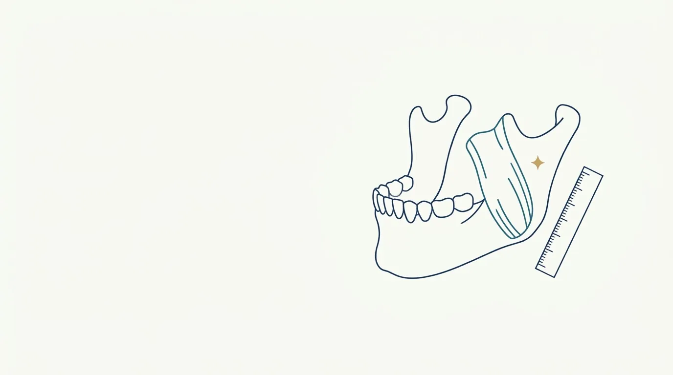
- Muscle
- Highlighted area
Quick facts
- Concern
- Square face, wide jaw angle, full lower face, sometimes pain or fatigue when chewing.
- Possible causes of the enlargement
- Muscle (masseter), bone (mandibular angle), gland (parotid), fat (subcutaneous or buccal), or a combination.
- Why it matters
- Toxin only acts on muscle. Treating without a diagnosis leads to frustration, asymmetry, or an unwanted effect.
- Simple test
- Palpate the masseter at rest and with the jaw clenched: if it hardens and bulges, there is a muscular component.
"I want to slim my face" is a phrase that often ends in botulinum toxin in the masseter. The reasoning seems obvious: wide face, large masseter, weaken the muscle. But the width of the lower face is not made of muscle alone. Bone, gland, and fat all contribute, and toxin acts on only one of them. Before treating, the professional's job is to find out what is actually creating the volume.
This page covers differential diagnosis, not injection technique. The treatment itself, with its risks and indications, is in botulinum toxin in the masseter, and chewing difficulties after treatment are in difficulty chewing after masseter treatment.
What makes up the width of the lower face?
The contour of the area is a sum. Understanding the layers helps locate what changed or what the patient wants to change.
| Component | Where it is | What it causes | Does toxin act? |
|---|---|---|---|
| Masseter muscle | From the zygomatic arch to the jaw angle | Enlargement of the lower face, mostly lateral, that grows when the teeth are clenched | Yes |
| Bone (mandibular angle, ramus) | Bony structure of the jaw | Wide or prominent angle, even at rest | No |
| Parotid gland | Over the masseter and behind the ramus | Diffuse enlargement, may be painful or fluctuate with eating | No |
| Subcutaneous fat | Over the muscle | Soft volume, unrelated to clenching | No |
| Deep cheek fat (Bichat's fat pad) | Between the masseter and the buccinator | Fullness of the middle and lower thirds | No; see deep medial cheek fat |
| Skin and laxity | Superficial | Look of heaviness and "jowls" | No |
In practice, there is almost always more than one component. The question is not "which one?" but "how much of each, and what can toxin change?".
How to examine?
The exam is simple, and the most useful information comes from palpation and from observation at rest and in motion.
- Observe the patient from the front, in profile, and at three-quarter view, at rest and with the teeth clenched. Side lighting highlights volumes; see lighting in the facial harmonization clinic.
- Palpate the masseter with your fingers over the area, asking the patient to clench and relax. A muscle that hardens and bulges under the fingers suggests a muscular component.
- Palpate the jaw angle at rest. A hard bony structure that does not change on clenching suggests a bony component.
- Assess the parotid region: diffuse enlargement, different consistency, tenderness, relation to meals.
- Assess the fat: pinching the skin and subcutaneous tissue over the area helps estimate thickness.
- Compare both sides: asymmetry is common and can guide the diagnosis (one side with hypertrophy, the other normal).
- Ask about habits: bruxism, clenching, one-sided chewing, chewing gum, headaches, and joint pain.
Which warning signs call for investigation?
Some findings fall outside the benign picture and call for referral or tests before any procedure:
- One-sided enlargement of recent onset, especially if rapid.
- Pain, localized hardening, or a palpable mass.
- Fever or inflammatory signs in the area.
- Changes in mouth opening (trismus) or limited jaw movement.
- Paresthesia or facial paralysis.
- Enlargement related to meals, which suggests a glandular condition.
- History of radiotherapy, surgery, or trauma in the area.
In these cases, the hypothesis is not aesthetic, and referral to a physician or an oral and maxillofacial surgeon is the priority. See patient referral in facial harmonization.
When to use imaging?
The clinical exam is enough in most cases of symmetric hypertrophy with a typical appearance. When in doubt, tests help:
- Ultrasound, useful for measuring muscle thickness and assessing the gland and fat. See ultrasound in facial harmonization.
- Panoramic X-ray or CT, which assess the bone of the angle and ramus and the relation to occlusion.
- Magnetic resonance imaging, when a mass is suspected or there is a relevant doubt.
The decision to order a test belongs to the professional, with clinical justification, and the result should be interpreted by someone competent to do so. In some practices, measuring muscle thickness before and after is a useful way to track the response.
What changes in management depending on the cause?
| Main hypothesis | Possible direction |
|---|---|
| Muscle hypertrophy | Consider toxin, explaining the gradual effect, risks, and maintenance |
| Bony component | Toxin does not solve it; discuss alternatives, including contouring surgery, and align expectations |
| Fat | Treatments for localized fat or fullness, or a review of the concern |
| Gland | Refer for medical evaluation |
| Combination | Treat the treatable component and explain the rest |
| Aesthetic concern without real enlargement | Reassess expectations and, at times, body dysmorphia; see body dysmorphic disorder in facial harmonization |
The key word is "explain". The patient needs to know that toxin thins the muscle, that the effect is gradual and temporary, that chewing strength may change, and that the final contour depends on what lies beneath.
What about the effect on the smile and chewing?
Even when the cause is muscular, toxin in the masseter can cause changes in chewing and in the smile, especially through spread to neighboring muscles such as the risorius and the zygomaticus. The topic is covered in difficulty chewing after masseter treatment and in the muscle map for botulinum toxin. The nearby nerve structure, such as the marginal mandibular branch, deserves anatomical knowledge.
How to talk to the patient
Document the findings and the reasoning. If the decision is to treat, record the specific consent. If not to treat, explain why and the alternatives. In both cases, standardized photos are part of the patient record; see standardized clinical photography.
And in Harmy?
Summary
Before toxin in the masseter, find out what creates the volume: muscle, bone, gland, fat, or a mix. The clinical exam at rest and in contraction, the history of habits, and, when needed, imaging guide the decision. Treat what is treatable, refer what falls outside the pattern, and make clear to the patient what toxin cannot change.
Frequently asked questions
How can I tell if my wide face is from the masseter?
The clinical exam helps: the masseter hardens and forms a bulge when you clench your teeth. If the enlargement is present at rest and does not change with clenching, it may be bone, fat, or gland. Imaging clears up doubts.
Does toxin in the masseter work for a wide face of bony origin?
No. Toxin acts on muscle and does not change bone. In cases of bony or fatty origin, the effect on contour is small or nonexistent, and a prior assessment avoids a wrong expectation.
Does bruxism always cause masseter hypertrophy?
Not always. Bruxism increases muscle activity and can lead to hypertrophy, but not every large masseter comes from bruxism, and not every case of bruxism causes visible enlargement.
When should I order imaging before toxin?
When the clinical exam is not conclusive, there is significant asymmetry, suspicion of a glandular or bony cause, pain, or rapid or one-sided enlargement. The decision is clinical and may include ultrasound or other tests, depending on the case.
Harmy images are illustrative simulations generated by AI from the patient's photo to support the conversation between practitioner and patient. They are not a promise of results. Indication, technique, product and volume are solely the decision of the licensed practitioner. This content is for information only and does not replace a clinical assessment.