
Assess the facial skeleton before injecting volume
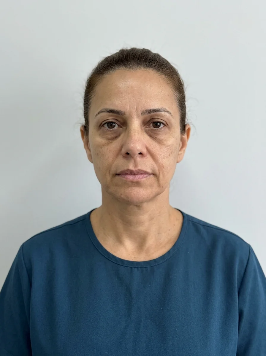
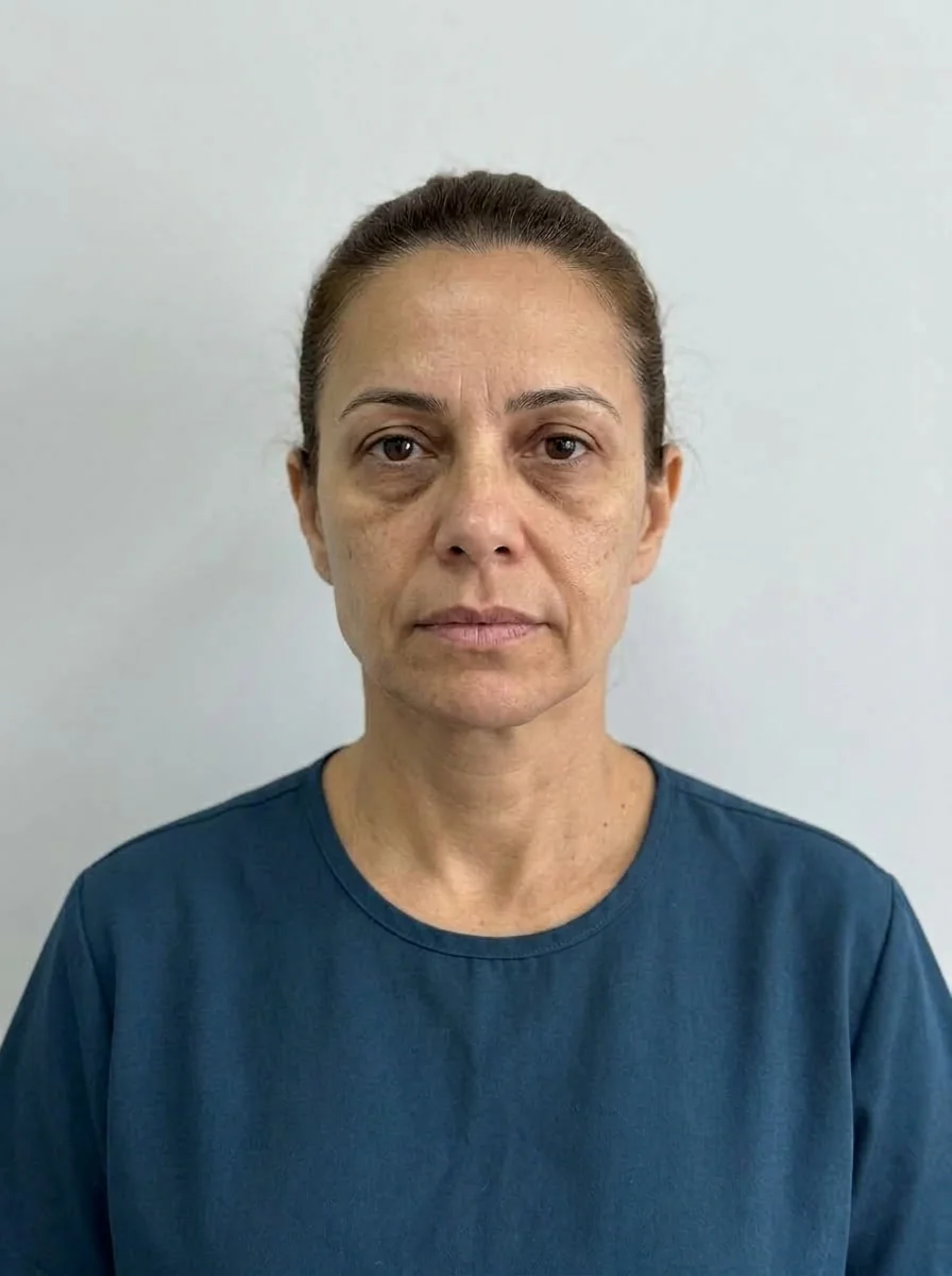 BeforeSimulation
- Highlighted area
Quick facts
- Topic
- Facial skeleton in the plan
- Central question
- Is the deficit in bone or in soft tissue?
- Warning sign
- A lot of volume for little effect
- Possible approach
- Treat, observe or refer
A patient arrives concerned about a "small" chin and asks for filler. Another wants to "enlarge the cheekbone" because the face looks flat. What is behind these requests? In some cases, loss of fat and soft tissue; in others, a skeleton with less projection that no reasonable amount of product can compensate for naturally. Telling the two apart is the first decision of a mature plan.
Why does the skeleton matter?
Bone is the framework on which fat, muscle and skin are arranged. Bony changes over time (facial bone resorption) alter the projection of the cheekbone, maxilla and mandible, and soft tissue descends or loses support. In young people, the skeleton also defines the base of the face: a recessed chin (retrogenia) or a poorly projected maxilla produce appearances that are not "lack of volume" in the soft tissue sense.
A plan without this reading usually falls into two errors: treating the symptom with ever-increasing volume, or promising what the procedure does not deliver. The overview by layers is in facial aging by layers.
What should you look for?
| Area | Skeletal question | Reference |
|---|---|---|
| Cheekbone | Has the projection of the zygomatic bone always been low? | Zygomatic bone |
| Midface | Is the maxilla poorly projected? | Maxilla and premaxilla |
| Chin | Is the chin recessed, or is the problem in the soft tissue? | Retrogenia |
| Mandible | Are the angle and body short, or is there laxity? | Mandible and mandibular angle |
| Profile | Is there convexity or retrusion that the front view hides? | Facial profile |
This table is a guide, not a diagnosis. Profile and angle analysis have their own pages: facial profile analysis: checklist, retrognathic profile: assessment and maxillary projection and midface in profile.
Which signs suggest the bone is the problem?
When several items are present, the conversation should cover the limits of filler. See when bone is the problem, not volume and chin and jaw: combining in the plan.
What can and cannot HOF do?
HOF can soften the perception of some deficits, as long as expectations are realistic and volumes are conservative. It does not change the skeleton. For significant deficits, the alternative may be orthodontics, orthognathic surgery or other surgeries, assessed by a licensed professional. A referral is not a failure of the plan: it is part of the care. See HOF integrated with dentistry and handing a plan over between professionals.
How do you talk to the patient?
Patients often think "volume is missing". Explaining that part of the face depends on bone changes the conversation, and it must be done without discouraging them or selling what cannot be delivered. See explaining what harmonization does not solve.
Does Harmy help here?
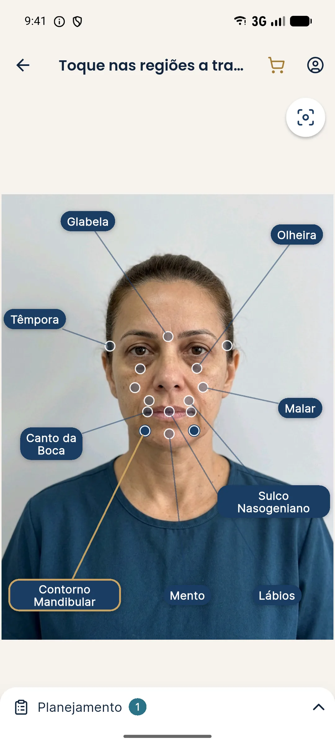
Harmy works with one front-facing photo and does not see profile or bony projection. It marks Cheekbone, Chin and Jawline, takes ml and intensity, and generates an illustrative projection. In cases of skeletal deficit, the projection may look better than the real result, because the AI does not account for the limit of soft tissue volume. So use the app with caution and make clear to the patient what it shows and what it does not. It is an illustrative simulation, not a promise of results. See limits of 2D simulation and when simulation gets in the way of the plan.
When should you request tests or a second opinion?
In most cases, clinical assessment is enough. But there are situations where an additional test or a second opinion helps you decide safely:
- Significant bone asymmetry, with functional or dental impact.
- A high-volume plan in a bony area, such as the chin or jaw, where the doubt between bone and soft tissue is large.
- A profile concern in a young patient with suspected significant retrusion.
- History of facial trauma or surgery that may have altered the skeleton.
- Pain or limited mouth opening, which call for joint assessment.
In these cases, referral to the dentist, orthodontist or surgeon is a care decision. Record the reason, the professional recommended and the guidance given to the patient. If they prefer to continue with HOF alone, document the explanation of the limits and their acceptance.
Another useful practice is to keep profile and 45-degree photos in the patient record, even though Harmy uses only the front view. These images help track changes and discuss the case with colleagues. See oblique facial analysis: three-quarter view and facial analysis in HOF step by step.
Finally, remember that teenagers and young adults may still have a skeleton in transformation. The decision to intervene should take this into account, and the approach with minors follows its own rules, as in saying no to a minor patient.
Summary
Before injecting, ask whether the deficit is in bone or in soft tissue. Examine from the front, in profile and at an angle, compare with older photos and be honest about limits. Refer when needed. The decision belongs to the licensed professional, after assessment, in line with the rules of their professional council, and the simulation is always illustrative.
Frequently asked questions
Why assess the skeleton before filling?
Because soft tissue volume depends on bone support. If the deficit is in the bone, filling soft tissue may take a lot of product, look artificial or fail to resolve the concern.
How do you tell a bone deficit from a soft tissue deficit?
By clinical assessment from the front, in profile and at an angle, palpation of the bony contour, comparison with older photos and, when needed, additional tests or referral.
Does filler replace surgery?
No. It can soften the perception of some deficits, but it does not correct skeletal changes. The decision about each route depends on the assessment and on what the patient wants.
Does Harmy consider the skeleton?
No. Harmy uses the front-facing photo and generates an illustrative projection of the marked areas. It does not assess bone, profile or bony projection.
Harmy images are illustrative simulations generated by AI from the patient's photo to support the conversation between practitioner and patient. They are not a promise of results. Indication, technique, product and volume are solely the decision of the licensed practitioner. This content is for information only and does not replace a clinical assessment.