
Maxillary Projection and the Midface in Profile: Assessment

- Highlighted area
Quick facts
- Topic
- The maxilla as support for the midface: nose, upper lip, fold and cheekbone depend on it
- Signs of under-projection
- Base of the nose seemingly lower, upper lip set back, deepened fold and flat cheekbone
- Tools
- Standardized profile photo, nasolabial and nasofacial angles, Ricketts and Steiner lines
- Caution
- A significant skeletal deficit calls for a specialist's assessment; volume is a complement, not a substitute
When you look at a person's profile, attention tends to go to the nose, the lips and the chin. The maxilla, which sits behind them, is rarely the star of the conversation, yet it supports much of what you see. An under-projected maxilla can make the nose look bigger, the upper lip set back and the fold deeper, without any of those structures being wrong on its own.
We will look at how to read that support, which signs point to under-projection and how it enters planning. The text is about assessment, at review level, and does not describe injection technique.
Why does the maxilla matter for the midface?
The maxilla forms the skeleton of the midface, from the nose to the upper lip. The premaxilla, the anterior part near the piriform aperture, supports the base of the nose and the upper lip. When that support is smaller, the soft tissues follow the bone and the relationship between nose and lip changes.
This explains why two patients with noses of the same size can have different profiles. In one, the maxilla projects and the nose looks proportionate. In the other, the maxilla sits further back and the same nose looks forward. This principle appears in the analysis of the whole in aesthetic profile: lips, nose and chin.
Which signs suggest maxillary under-projection?
No single sign proves it, but the set points toward investigating:
- Nasolabial angle very closed or very open compared with what is expected for the patient, per the nasolabial angle.
- Upper lip set back relative to the profile reference line, such as the Steiner S line or the Ricketts line.
- Deep, early nasolabial fold, with a flat cheekbone.
- Flattened paranasal area, seen in profile and three-quarter view.
- Nose that looks "drooping" or bigger without the cartilage being the cause.
- Complaint of a tired midface with no obvious soft tissue volume loss.
Patients with a different bone pattern or ancestry have different references, and comparing them with the "textbook standard" can lead to error. The caution is in respecting ethnic features in the facial harmonization plan.
How to photograph and measure the profile?
The profile photo requires a standard: head in the Frankfurt plane, neutral expression, relaxed lips, hair pulled back and no accessories. Any head tilt changes every measurement. Detailed guidance is in profile facial analysis: checklist and in hair, earrings and glasses in the assessment photo.
| Measurement | What it tells you | Note |
|---|---|---|
| Nasolabial angle | Relationship between the columella and the upper lip | Depends on tip rotation and lip position |
| Nasofacial angle | Slope of the nasal dorsum relative to the facial plane | Helps tell a large nose from small support |
| Distance from the subnasale to a reference vertical | Projection of the base of the nose and the lip | Compare with the chin and forehead |
| Facial convexity | Overall profile | See facial convexity angle |
Reference values vary by author, sex and population, so they are left off this page. What counts is the reading as a whole, done by the professional who examines the patient.
What can volume do, and not do, for the maxilla?
When the deficit is mild to moderate and mainly in soft tissue, adding volume in the paranasal area and around the cheekbone can improve the look of support and soften the set-back appearance. This is a decision for the qualified professional, after assessment, with attention to the arteries of the area, which are high-risk, such as the angular artery and the infraorbital artery. Hyaluronidase must be on hand when the product is hyaluronic acid.
When the deficit is significantly skeletal, the answer is different. Orthognathic surgery or orthodontic treatment are the described routes to correct the bone, and volume is a complement. The distinction is the same as in when bone is the problem, not volume.
How does the maxilla relate to the cheekbone, lip and fold?
The cheekbone and the maxilla share the lateral transition of the midface, and the malar projection described in ideal malar projection acts on part of the same support. The nasolabial fold, when deep, is sometimes a symptom of insufficient support and not only of local loss, and that is the logic of treating the fold or the cause of the fold.
The upper lip is directly influenced by the premaxilla: the vermilion and the philtrum depend on the support. The relationship is considered in nose and upper lip: combining them in the plan. The whole picture is in complete midface in the plan, and the nose and cheekbone areas have their own pages.
How to talk about this with the patient without alarming them?
Most patients do not know the maxilla and worry about the nose. Showing the profile photo and drawing a reference line is clearer than any technical term. Be careful with promises: suggesting that volume will "fix the nose" is inappropriate. What can be said is that support changes how the whole reads and that the assessment is individual.
For the conversation about nonsurgical rhinoplasty and expectations, see explaining nonsurgical rhinoplasty to the patient and explaining what facial harmonization does not solve.
Frequently asked questions
What is maxillary projection in profile?
It is how far the maxilla and the premaxilla area advance in profile. It serves as the base for the tip and base of the nose, the upper lip and part of the midface.
Does an under-projected maxilla change the nose?
It can change how the nose reads. With less support, the base of the nose and the upper lip look set back, and the nose may look bigger or more drooping than it really is.
Does filler correct maxillary under-projection?
It can improve the look of support in moderate deficits, at the professional's discretion. In a significant skeletal deficit, an orthodontic or surgical assessment is needed, and volume is a complement.
Does Harmy assess the maxilla?
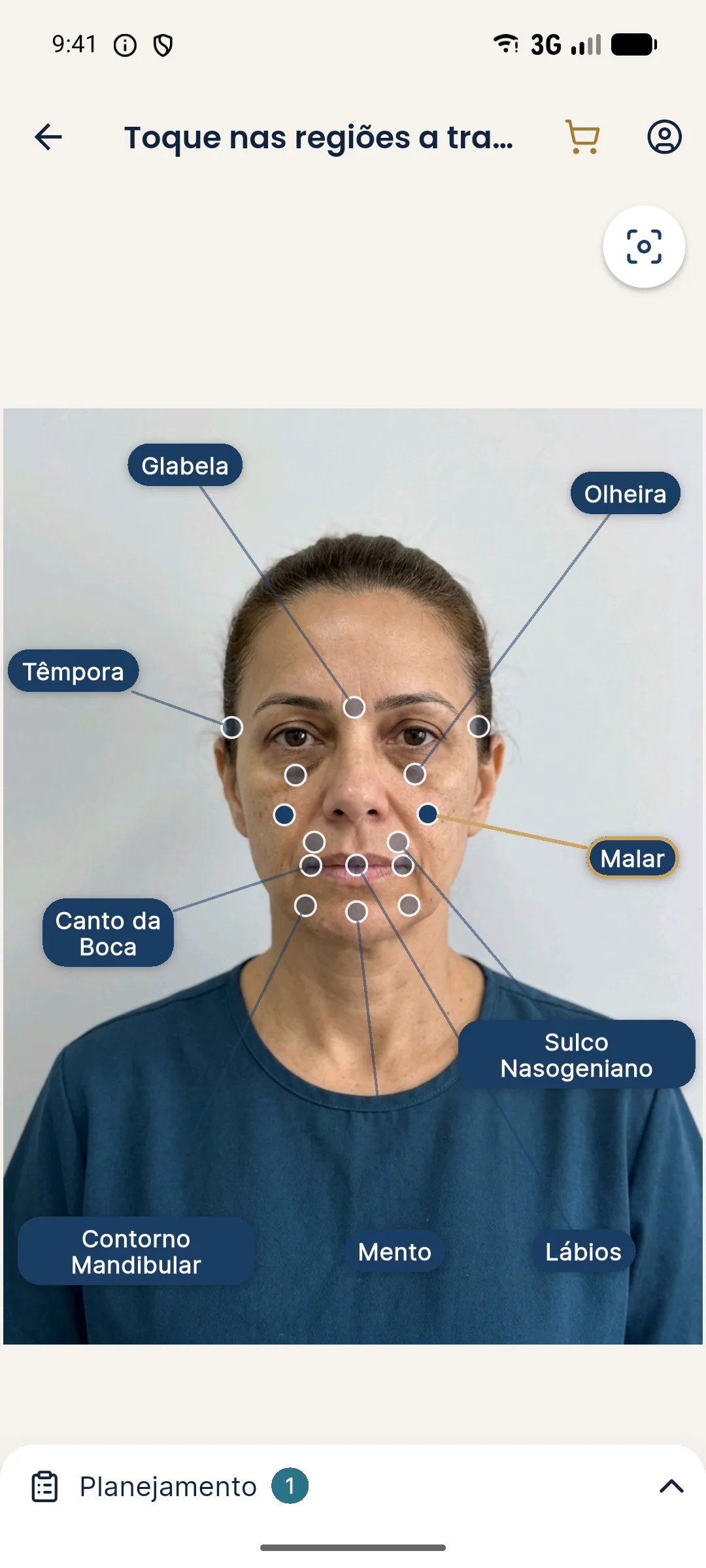
No. The app uses only the frontal photo. Reading maxillary projection is a clinical task, done in profile, with standardized photos and an exam.
Harmy images are illustrative simulations generated by AI from the patient's photo to support the conversation between practitioner and patient. They are not a promise of results. Indication, technique, product and volume are solely the decision of the licensed practitioner. This content is for information only and does not replace a clinical assessment.