
Malar Region (Cheekbone): Anatomy and Filler
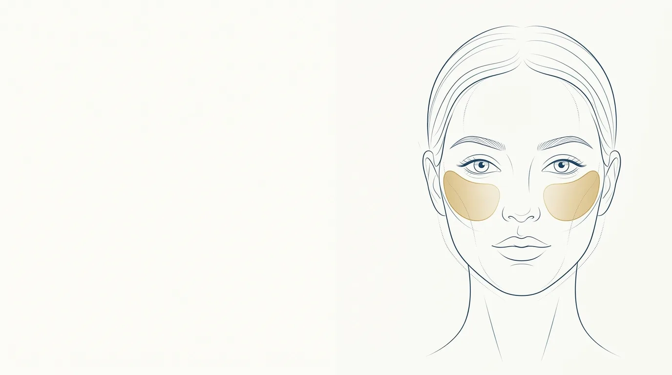
The malar region is the cheekbone, the most projected part of the midface in frontal view. It works as a supporting structure: when it loses volume, the midface descends, the tear trough becomes more marked, and the nasolabial fold deepens. That is why the malar area is, in many plans, the starting point of treatment.
What are the anatomical boundaries of the malar region?
The malar region lies over the body of the zygomatic bone and the adjacent portion of the maxilla. The practical boundaries are:
- Superior: the inferior orbital rim and the transition to the lower eyelid.
- Medial: the paranasal region and the start of the nasolabial fold.
- Lateral: the continuity with the zygomatic arch.
- Inferior: the cheek and the mid-cheek groove.
Among the important structures are the fat compartments of the midface (superficial and deep, such as the malar fat pad and the suborbicularis oculi fat), the zygomatic retaining ligaments, and the zygomaticus major and minor and levator labii superioris muscles, which take part in the smile.
Vessels and nerves to watch
- Infraorbital artery and nerve, which emerge through the infraorbital foramen, below the rim at the mid-pupillary line.
- Facial artery and angular artery, at the medial transition with the nose and the fold.
- Transverse facial artery and zygomaticofacial branches in the lateral portion.
- Zygomatic and buccal branches of the facial nerve, which supply the smile musculature.
The medial portion of the malar area, near the angular artery and the infraorbital foramen, calls for more caution than the lateral portion.
What do patients usually ask for?
Patients rarely say "malar". Their concerns sound like this:
- "My face has dropped."
- "I want more defined cheekbones."
- "I look tired and my face looks deflated."
- "I want to lift my face without surgery."
Assess the patient from the front, in three-quarter view, and in profile. The projection of the cheekbone, the position of its highest point, and the relationship with the tear trough and the fold guide the indication. In faces that are already wide, enlarging the malar area can make the face look heavier, and the better option may be to work on other areas.
Which procedures can be done on the malar area?
The main one is malar filler with higher G' hyaluronic acid, suited to providing support. Some professionals use calcium hydroxyapatite or other collagen biostimulators aiming for gradual stimulation and support.
The malar area is usually planned together with:
- Zygomatic filler, for the lateral portion.
- Tear trough filler, since malar support can reduce the product needed in the groove.
- Nasolabial fold filler, which usually needs less volume once the midface has been treated.
What are the usual volumes for the malar area?
Usual ranges in ml of hyaluronic acid, at the discretion of the licensed professional, after assessment:
| Situation | Usual range per side | Note |
|---|---|---|
| Subtle enhancement | 0.5 to 1 ml | Young patient, focus on contour |
| Moderate loss of support | 1 to 1.5 ml | Most common case |
| Marked loss | 1.5 to 2 ml or more, in sessions | Splitting helps assess the response |
| Durability | Generally 12 to 18 months | Varies with product and patient |
In patients with significant loss, treating in stages helps avoid excess volume and an artificial look.
Care, contraindications, and complications
General contraindications: active infection or skin lesion at the site, pregnancy and breastfeeding, uncontrolled autoimmune disease, allergy to the components, and unrealistic expectations. Record the use of anticoagulants and antiplatelet agents.
Reported complications:
- Swelling and bruising in the first few days.
- Excess volume, giving a "puffy" face.
- Asymmetry between the sides.
- Palpable lumps or irregularities.
- Vascular occlusion, more likely in the medial portion, with a risk of necrosis.
When injecting hyaluronic acid in the malar area, keep hyaluronidase available in the room. More guidance in safety and complications.
How to assess loss of support in the midface?
The malar area is one of the regions where the assessment changes the plan the most. Some practical points:
- Three-quarter view: this is where the loss of cheekbone projection shows up most clearly.
- Point of greatest projection: in young faces it tends to sit higher and more lateral; with aging, the volume tends to descend.
- Relationship with the tear trough: if the step between eyelid and cheek is increased, malar support can help.
- Relationship with the nasolabial fold: a deep fold with a flat midface usually indicates the cause lies above it.
- Face width: faces that are already wide may call for more caution with lateral volume.
Assessing the face by thirds helps you see the midface within the proportions of the whole face. Standardized clinical photography lets you compare before and after under the same light.
How does the malar area appear in Harmy?
In Harmy, the area is the Cheekbone point in the marking step (malar, on this site). The same point also represents zygomatic filler in the app's catalog. After the frontal photo, the app detects the face on the device and shows the points with labels; tap Cheekbone to include it in the plan.
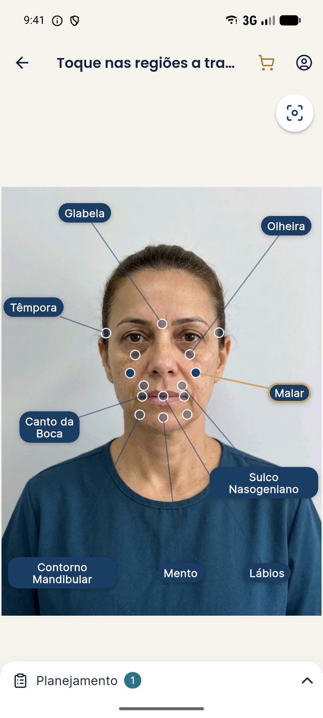
In the Plan, set the volume in ml (0.5 ml steps, from 0.5 to 5 ml). Because it is a bilateral area, the value is the total, split between both sides. The simulation on this site used 2 ml in total. The projection changes only what was marked, with no skin retouching, makeup, or change of expression.
See the step by step in marking areas and volumes.
How to talk to the patient about the malar area
Because the malar area treats the cause of concerns that the patient attributes to other areas, it is worth showing the effect on their photo. An illustrative simulation of the malar area, compared with the original photo, helps the patient understand why you propose starting with the cheekbone and not with the smile lines.
Present the projection as a support tool and not as a promise. To build the proposal from the volume, see pricing per ml.
Frequently asked questions
What is the difference between malar and zygomatic?
In practice, malar usually refers to the anterior, more central portion of the cheekbone, and zygomatic to the lateral portion, along the arch. Both areas are treated in an integrated way in the midface.
How much filler is used in the malar area?
Generally 0.5 to 2 ml per side, at the discretion of the licensed professional after assessment. Larger volume losses are often treated over more than one session.
Does filling the malar area improve smile lines?
It can help. Restoring midface support usually softens the nasolabial fold and the tear trough indirectly, but the effect varies from patient to patient.
How long does malar filler last?
Generally 12 to 18 months, depending on the product, the volume, and the patient's metabolism.
How do I simulate the malar area in Harmy?
Mark the Cheekbone point on the frontal photo and set the volume in ml in the Plan. The value is the total, split between both sides.
Harmy images are illustrative simulations generated by AI from the patient's photo to support the conversation between practitioner and patient. They are not a promise of results. Indication, technique, product and volume are solely the decision of the licensed practitioner. This content is for information only and does not replace a clinical assessment.