
Clinical photography in facial harmonization
A good photo is clinical data. Practical guides to shoot with your phone, keep photos consistent between visits, compare results and store images securely.
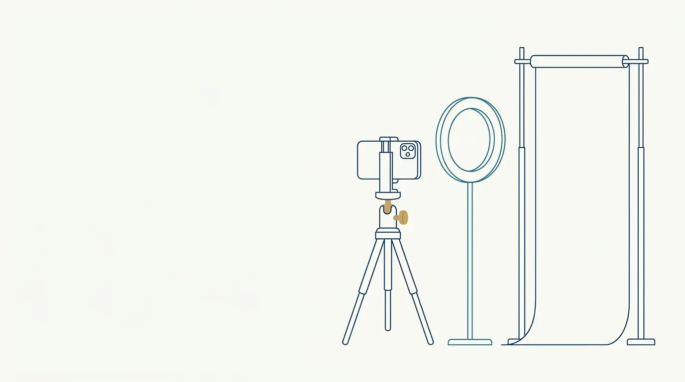
 2D photography or 3D scanner in the facial harmonization clinicWhen a 2D phone is enough and when a 3D scanner makes a difference in a facial harmonization clinic: cost, learning curve, standardization, what each measures.
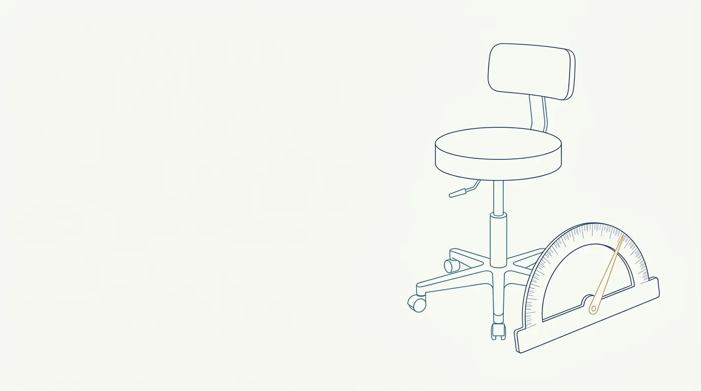 45-degree face photo: how to standardize the angleHow to get the 45-degree oblique view of the face in a repeatable way, what it shows that the frontal view hides and how to mark the position in the clinic.
 A Forced Smile Misleads the Assessment: How to Avoid ItThe difference between a posed and a spontaneous smile in clinical photos, what each one distorts and how to get a faithful record of the smile.
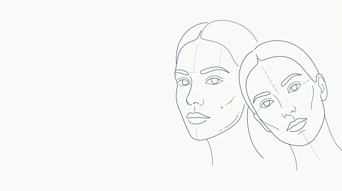 A Tilted Face in the Photo Changes Perceived SymmetryHow head rotation and tilt change how symmetry reads in clinical photos, and how to correct posture before the shot.
 After photo looks worse than the before: what happenedWhy the after photo can look worse than the before even with a good result: light, angle, expression, swelling and how to explain it to the patient.
 Aligning Photos by the Eyes to Compare Before and AfterHow to use the pupils as a scale and rotation reference to overlay before and after photos without misleading the reading of the result.
 Background for clinical photos: color, distance, neutralityWhich background to use in clinical photos: neutral color, distance from the wall to avoid shadow, and how it changes perceived skin tone and exposure.
 Backing Up Clinical Photos: A Simple RoutineHow to apply the 3-2-1 rule to backing up patient photos, with separate copies, encryption, restore testing and periodic review.
 Bad photo for AI projection: what gets in the wayShadow, angle, expression, filters and low sharpness degrade AI projection. What to avoid in the photo and how to spot the problem before spending a projection.
 Bite and Teeth in the Lip Photo: Jaw PositionHow to position the jaw and teeth in the lip photo: soft occlusion, relaxed lips and no clenching, to record lip volume faithfully.
 Bruise and swelling photo for tracking progressHow to document bruising and swelling with daily photos, a reference ruler and constant light, to track progress and guide the patient.
 Camera Height for Patient Photos: Level With the EyesWhy the camera should sit at the patient's eye level and how a high or low camera changes the chin, nose and folds in the clinical photo.
 Chin profile photo for planningHow to photograph the chin profile with a vertical reference line, neutral chin, and Frankfort plane to plan projection and record progress.
 Clinical photo resolution and format: HEIC or JPEGWhich resolution and format to use for clinical face photos: HEIC, JPEG or PNG, file size, compression and compatibility with the patient record.
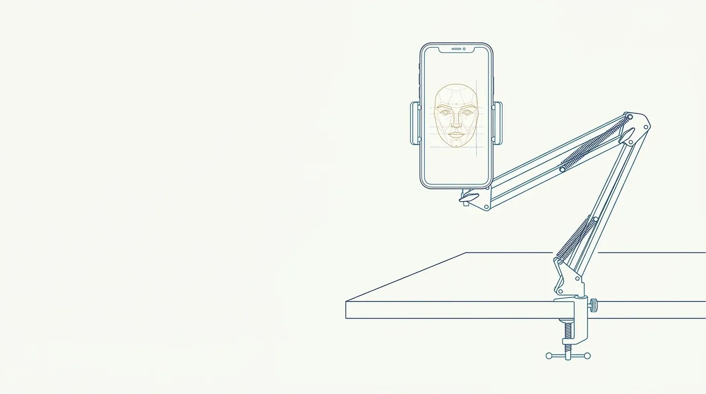 Clinical Photos With a Phone: How to Take Quality ShotsPhone setup for patient photos without accessories: camera, resolution, grid, locks, light, distance and a routine that gives comparable photos.
 Clinical video of the patient: when to recordWhen clinical video adds information beyond the photo: facial dynamics, speech and smile. What to record, how to standardize it and consent precautions.
 Cloud storage for patient photos: what to requireBefore storing patient photos in the cloud, check server location, encryption, access control, logs and data exit. A checklist for the clinic.
 Common Mistakes in Before and After PhotosA list of photography mistakes that mislead the reading of results in HOF before and afters: light, angle, expression, swelling, editing and how to avoid each.
 Complication photo: how to record it safelyWhat to show, when to photograph and what to note in the chart when recording a complication, with patient care always the priority.
 Consent for Clinical Video and StorageClinical video captures the patient's image, voice and movement. See what the consent should cover, how to store it and when to ask for separate authorization.
 Correcting Brightness and Contrast in a Clinical PhotoHow to adjust brightness and contrast of clinical photos equally in both images, without altering the result, and when it is better not to edit.
 Covering the Eyes in a Patient Photo: Does It Protect?Does an eye bar guarantee anonymity? See why covering the eyes does not anonymize the patient and which precautions apply to the clinical image.
 Cropping and rotating clinical photos: what is allowedCropping, rotating and straightening a clinical photo: what you can do without hiding information, what to avoid, and how to keep the original.
 Different Skin Tone Between Photos: CausesWhy skin tone changes from one clinical photo to another: tan, light, white balance and camera, and how to separate what belongs to the patient.
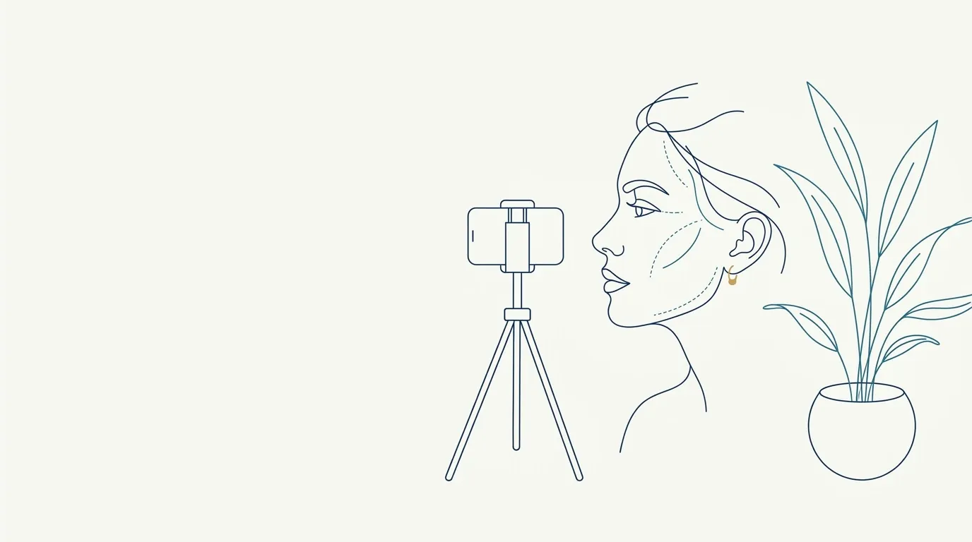 Dynamic photos for botulinum toxin: expressions to recordWhich expressions to photograph before botulinum toxin: forehead, glabella and eyes at rest and in contraction, with standard prompts and labels for follow-up.
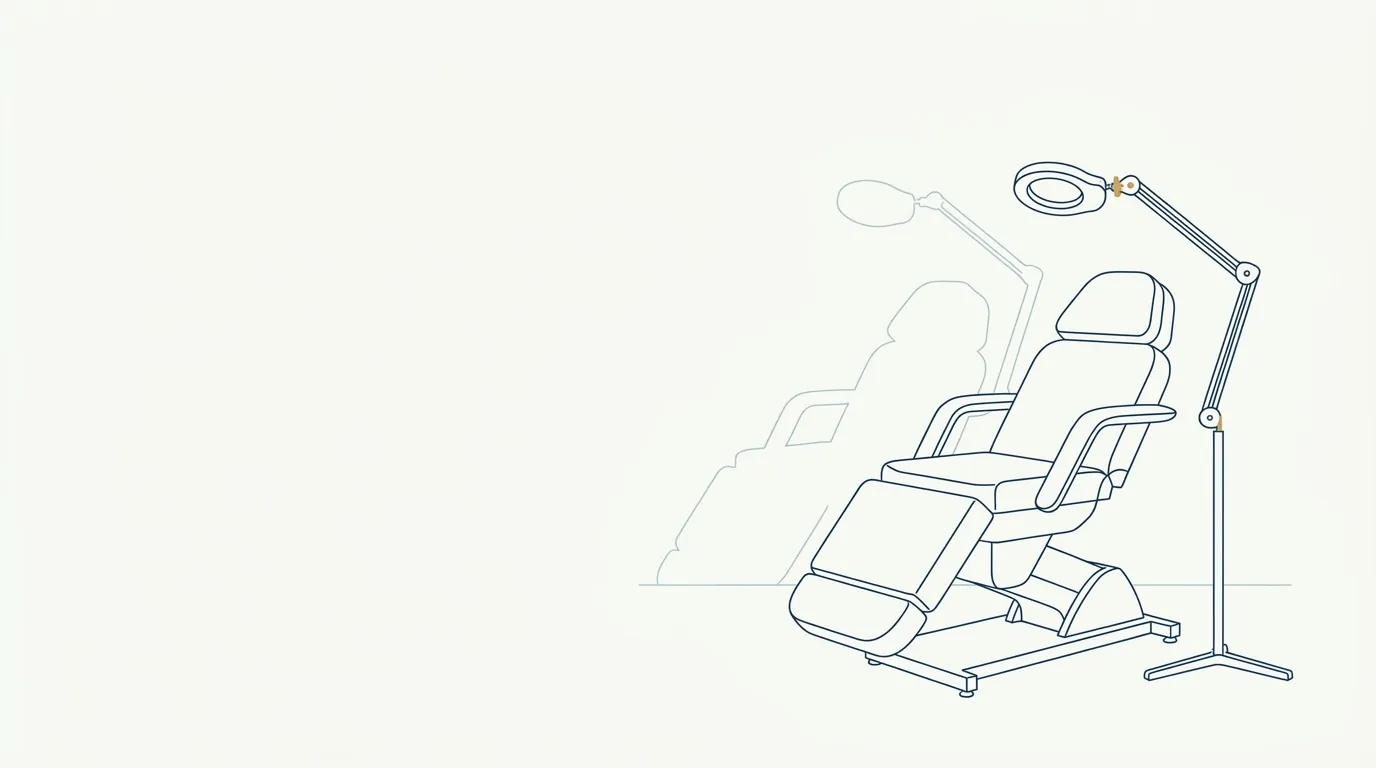 Facial shadows in clinical photos: causes and fixesLight from above, below or the side casts shadows that change tear troughs, folds and chin in clinical photos. Learn the causes and how to fix each one.
 Forehead photo with raised eyebrows for documentationHow to record frontalis contraction with raised eyebrows to document forehead lines and brow position before and after botulinum toxin.
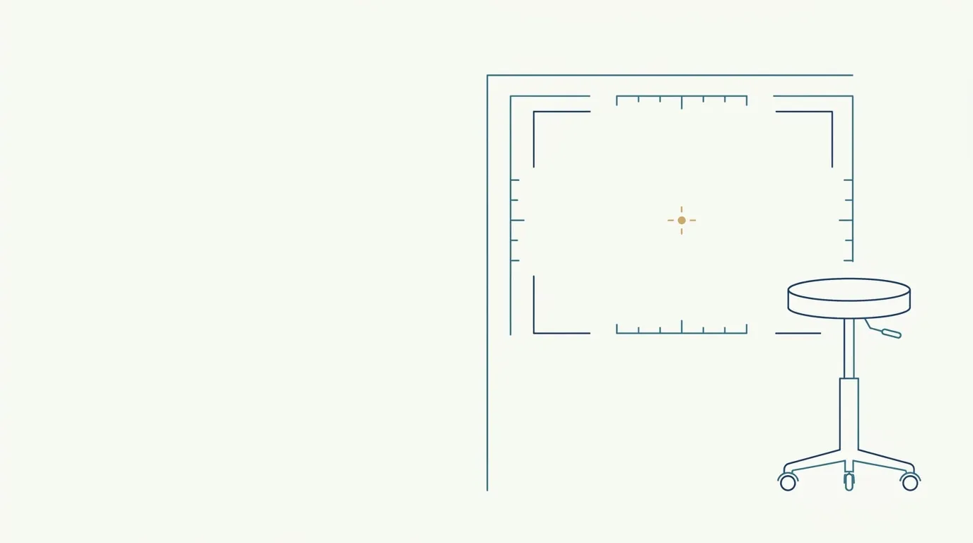 Framing the Patient's Front-Facing Photo Step by StepHow to frame the patient's front-facing photo from the top of the head to the neck, with the face centered, level and in the same proportion at every visit.
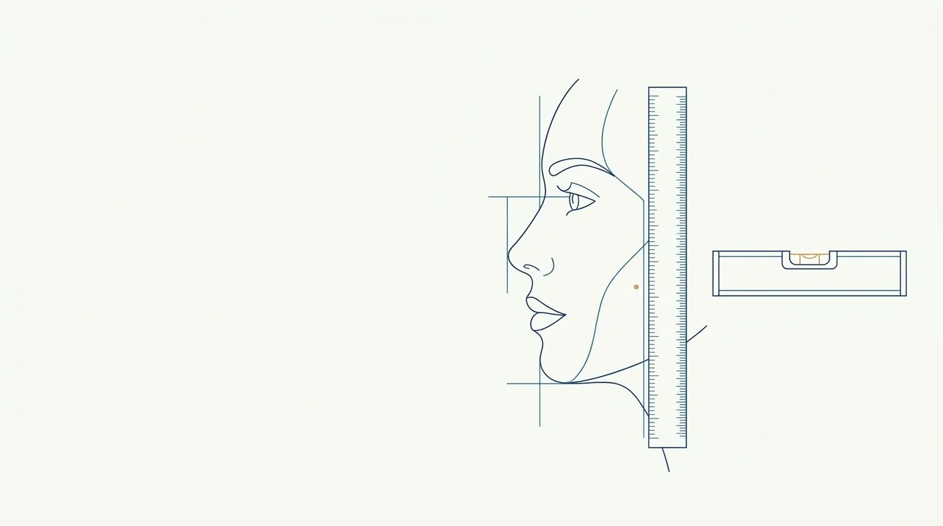 Frankfurt plane in the clinical photo: positioning the headWhat the Frankfurt plane is, how to align the patient's head before taking the clinical photo and how to check level without expensive instruments.
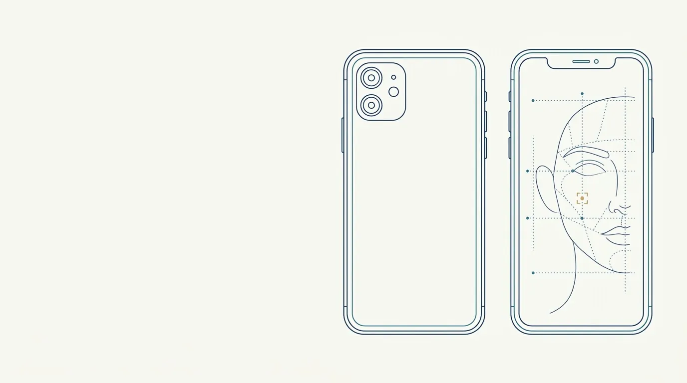 Front or Rear Camera for the Clinical PhotoFront (selfie) or rear camera for photographing patients: quality, mirror effect and distortion, and which to choose in the facial harmonization routine.
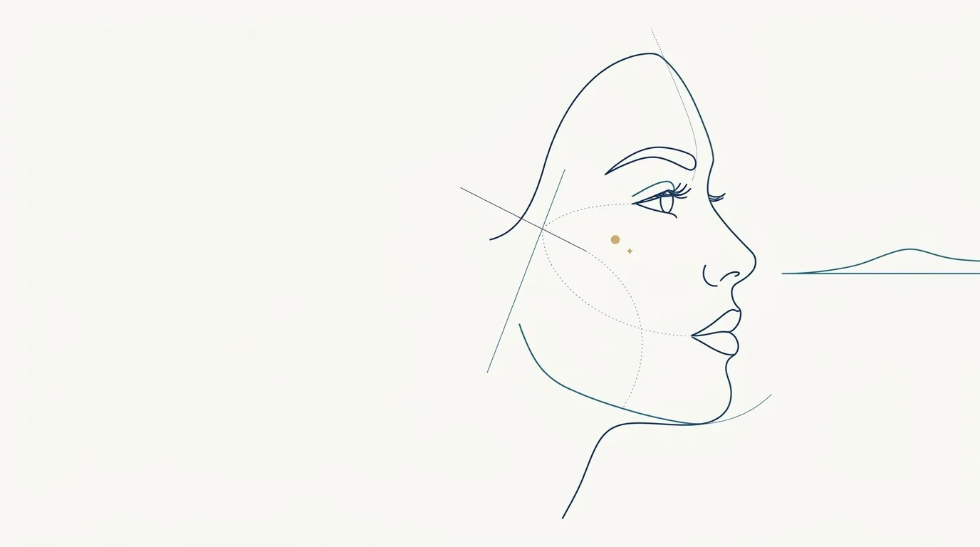 Gaze at the horizon in the clinical photoWhere the patient should look in a clinical photo: gaze direction, natural head position and how to avoid a raised or tucked chin.
 Glabella Photo in Contraction to Document Botulinum ToxinHow to photograph the glabella at rest and in contraction (angry expression) to document botulinum toxin before and at follow-up, with a repeatable standard.
 Glasses, Earrings and Accessories in the Clinical PhotoWhich accessories to remove before the clinical face photo, why, and how to record what cannot be removed so the comparison is not distorted.
 Hair Pulled Back in the Clinical Photo: What Must ShowWhy to pull hair back for the clinical photo: forehead, ears and hairline visible, with guidance for long hair, bangs and short hair.
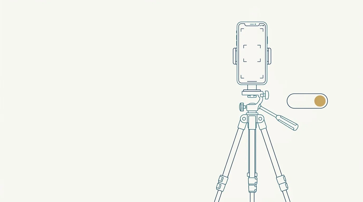 HDR and portrait mode in clinical photos: turn them off?HDR, portrait mode and automatic beautification alter edges, texture and shadows. What to turn off on the camera for a faithful clinical photo.
 How long to keep photos in the patient recordRetention period for clinical photos in the patient record, safe disposal and deletion at the patient's request: what to check in law, board rules and LGPD.
 How to name clinical photo filesA file-naming standard for clinical photos that keeps the patient chart organized without exposing the patient's name. Examples, field table and checklist.
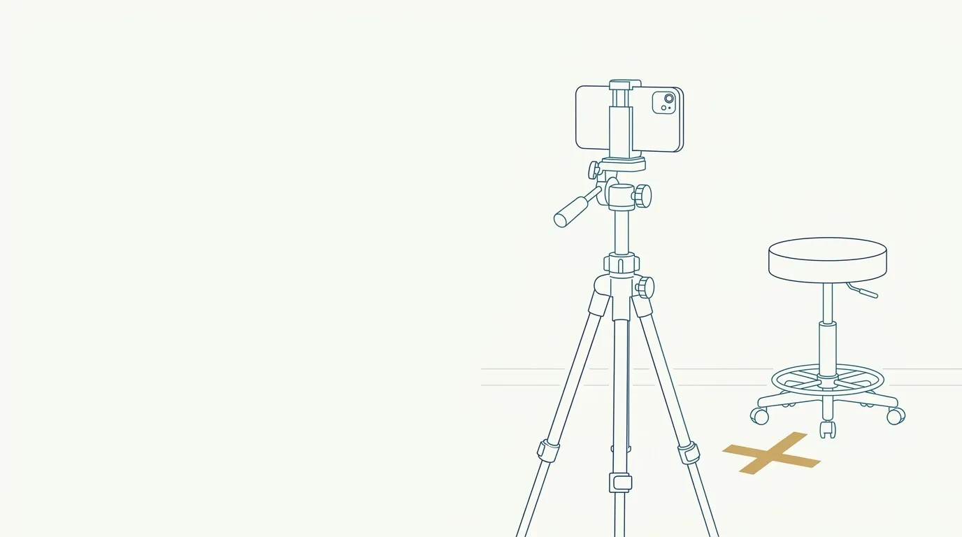 How to standardize photos between HOF visitsA setup sheet for clinical photos to repeat light, distance, height and position at every visit and compare before and after with confidence.
 Image Consent for Clinical DocumentationA chart photo and a publicity photo are different things. See how to handle image consent for clinical documentation in orofacial harmonization.
 Light for clinical facial photos: natural, diffuse or flashHow to position light in a clinical face photo so it does not fake volume or volume loss: natural light, diffuse light, ring light and flash.
 Lip photos for documentation: rest, smile and profileA short, repeatable sequence for photographing the lips: rest, smile, profile and speech, with lighting, distance and patient prep for a fair comparison.
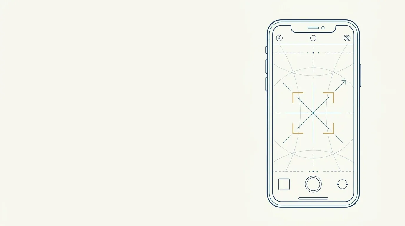 Locking Exposure and Focus on Your Phone for Clinical PhotosHow to use AE/AF lock on your phone to fix exposure and focus, keep brightness consistent between photos and stop the camera changing with every shot.
 Makeup in clinical photos: why ask for a clean faceFoundation, concealer, contouring and lipstick change skin and proportions in clinical photos. How to ask for a clean face and compare without bias.
 Marking floor and wall positions for clinical photosHow to use floor tape, a wall mark and a fixed height so the patient stands in the same spot in every clinical photo, with a setup checklist.
 Masseter and jawline photos: useful anglesHow to photograph the masseter and jawline in frontal, oblique, and underneath views, with the right light, to assess lateral volume and track progress.
 Metadata and location in patient photosEXIF, GPS, date and device model travel inside the photo. See what metadata reveals, when to strip it and how to avoid leaking patient data.
 Neck and double chin photos in the assessmentHow to photograph the neck and double chin with a neutral chin, head in Frankfort, and profile, without hiding or inventing submental fat or platysmal bands.
 Neutral Expression in Clinical Photos: How to Ask the PatientA script for getting a relaxed mouth, steady gaze and smooth forehead in clinical photos, with common mistakes and how to standardize across visits.
 Nodule photo for clinical follow-upHow to photograph a nodule with raking light to show relief, a scale ruler and a defined frequency, to track progress with a standard.
 Nose photos for nonsurgical rhinoplasty: frontal, profile, baseWhich nose photos to take before nonsurgical rhinoplasty: frontal, profile, obliques, and base, with distance, light, and errors that distort the assessment.
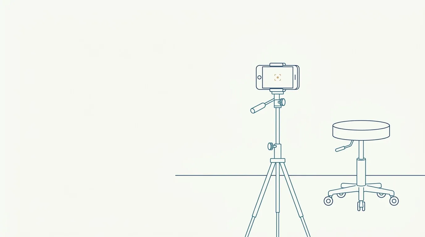 One frontal photo for planning: what it needs to haveMinimum requirements for the single frontal photo Harmy uses to detect the face and project: framing, light, expression, resolution and what ruins it.
 Patient Asks You to Delete Their Photo: How to RespondWhat to do when a patient asks you to delete their photos: the LGPD right to erasure, chart retention, marketing photos and backups.
 Patient photos in a lecture or congress: precautionsWhat to do before projecting a patient photo in a lecture, congress, or course: specific authorization, real anonymization, covering identifiers, file control.
 Patient photos on personal WhatsApp: risksThe risks of storing or sending patient photos on personal WhatsApp: automatic backup, camera roll, loss of control, and safer alternatives.
 Patient photos on social media: legal risksThe risks of posting patient photos on social media, in plain terms: CFO, CFM, LGPD, consent and what to check before publicizing.
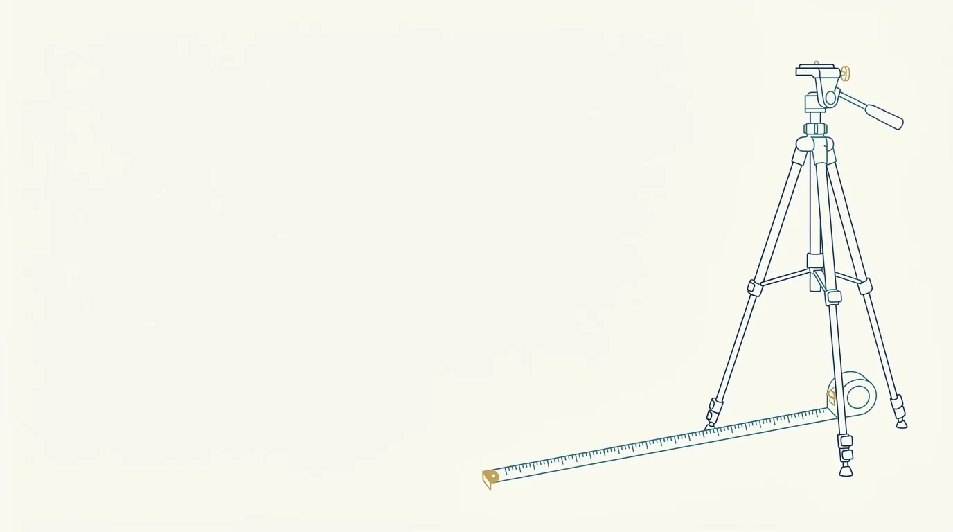 Phone Distance in the Patient Photo: How Many MetersWhat distance to use between the phone and the patient in a clinical photo, how to fix it with floor marks and how it relates to facial distortion.
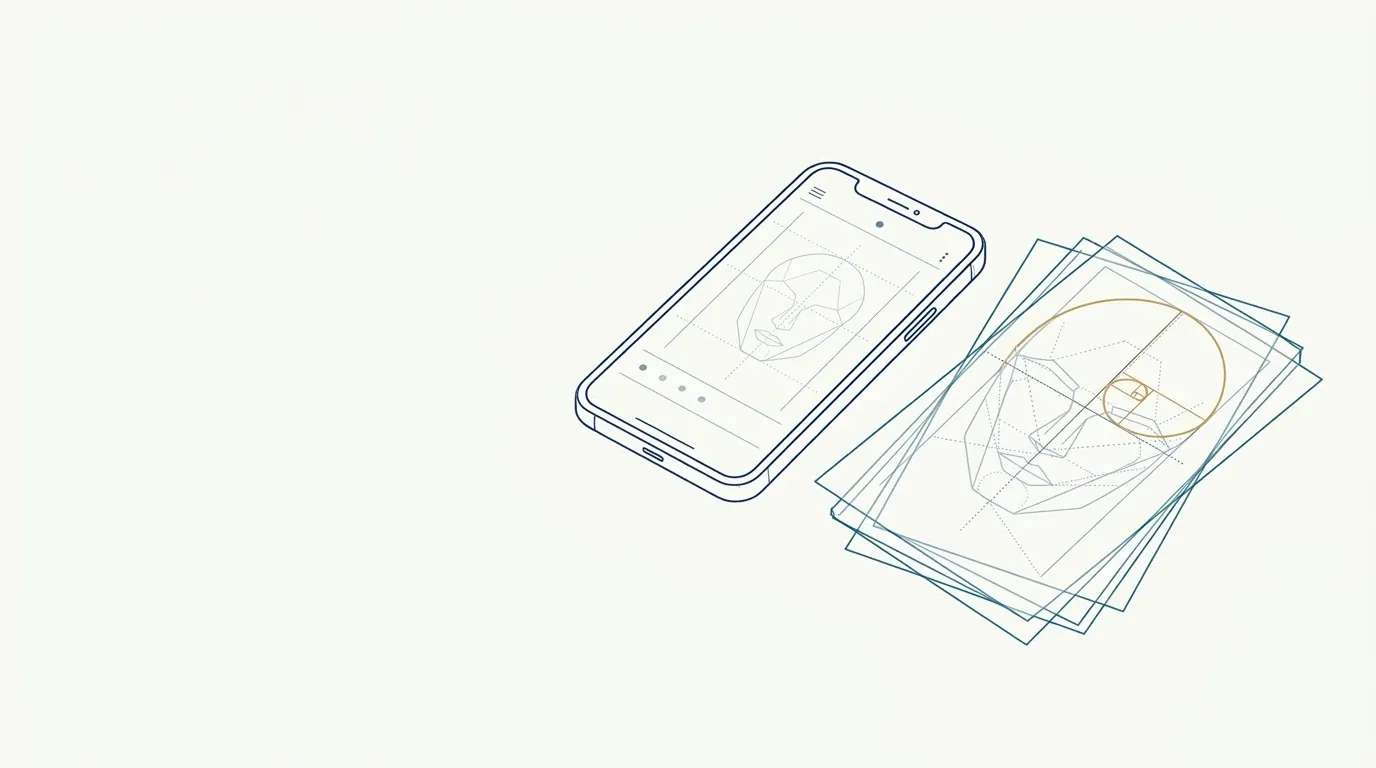 Phone Filters and Beauty Mode in Clinical PhotosHow to turn off skin smoothing, beauty mode and aggressive HDR on your camera so the clinical photo shows the real face and can be compared.
 Phone Flash in Clinical Photos: Why to Avoid ItPhone flash flattens relief, blows out skin shine and distorts color. Here is why to avoid it and which lighting alternatives to use for clinical photos.
 Phone gallery and patient photos: keep them separateWhy patient photos should not sit in your personal phone gallery: cloud sync, accidental sharing, backups, and how to keep them separate.
 Phone zoom in clinical photos: use it or step backOptical zoom, digital zoom, and phone lenses in clinical photos: when to use them, why moving closer or farther changes the face, and how to keep quality.
 Photo App for an Aesthetic Clinic: What to RequirePrivacy and standardization criteria for choosing a clinical photo app: access, cloud, sharing, export and deletion.
 Photo of ischemia signs for the chartHow to record pallor, livedo and other ischemia signs with a timestamp in the chart, without delaying care or the emergency response.
 Photo With a Ruler to Measure a Nodule or LesionHow to photograph a nodule or lesion with a visible scale ruler and a perpendicular angle to track its size across visits.
 Photo with a tattoo or mark that identifies the patientTattoos, scars, moles and piercings identify a patient even without the eyes. How to cover or crop for publication and what to keep in the chart.
 Photographing crow's feet in a forced smileHow to record the lines at the corners of the eyes at rest, in a natural and a forced smile, showing the orbicularis pattern without exaggerating them.
 Photographing every session in staged treatmentsHow to build a photo timeline in staged treatments: what to record at each session, when, and how to keep the same standard across sessions.
 Photographing the gummy smile for clinical documentationHow to document a gummy smile: resting and maximum-smile photos, without forcing, measuring gum exposure with repeatable framing and lighting.
 Photographing the midface to assess cheekbone volumeHow to photograph the midface to assess cheekbone volume: side and oblique light to read projection shadows without mistaking shadow for volume loss.
 Photos for comparing the result of a procedureThe minimum sequence for a fair before and after comparison: what to repeat, when to photograph again and how to label so the result is not misleading.
 Photos of minor patients: authorizationHow to handle clinical photos of minor patients: legal guardian authorization, extra caution, the minor's assent, and limits on use and publication.
 Practice lighting and skin tone in photosHow the color temperature of lamps changes skin tone in clinical photos and how to keep it consistent across rooms, times of day and visits.
 Profile photo of the face: how to take it in the clinicHow to photograph a true 90-degree profile, with bony landmarks and the Frankfort plane, to assess the nose, chin, and jawline.
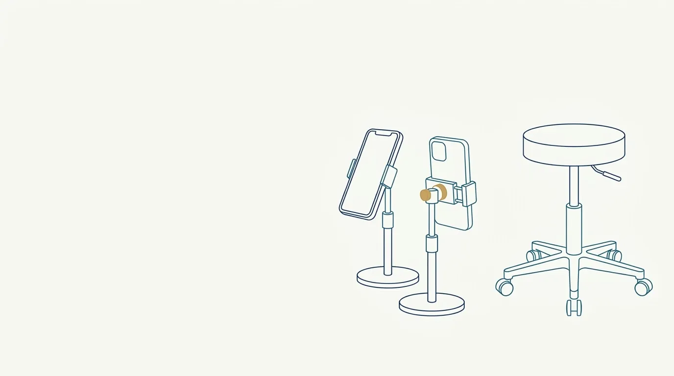 Resting photo vs dynamic photo: what is the differenceA resting photo shows the face without tension; a dynamic one shows it in action. When to use each, what to ask the patient and how to label the record.
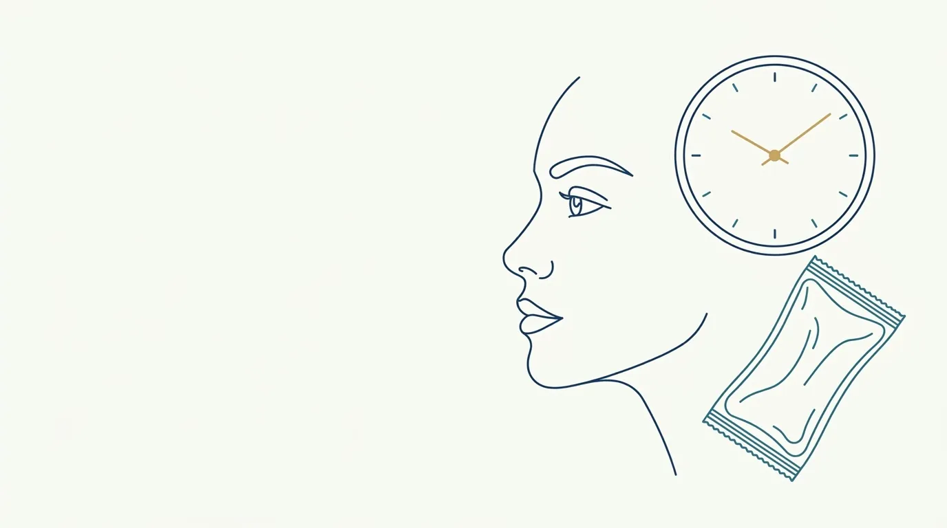 Result photos while swelling is still presentWhy an early photo misleads: swelling, bruising, and position change the face. When to photograph, how to label, and what to tell a patient who wants results.
 Ring light in clinical photos: advantages and pitfallsA ring light gives even frontal light but creates glare in the eyes and skin and erases useful shadows. When to use it in clinical photos and how to adjust it.
 Sending a Patient Photo to the Manufacturer or Tech SupportHow to send a patient photo to the manufacturer or technical support after a complication: restricted purpose, anonymization and a chart record.
 Separate Consent to Publish a Patient's ImageWhy to use two distinct forms, one for treatment and another for publishing images, and how to handle the patient's revocation of consent.
 Sharing a Patient Photo With Another ProfessionalHow to send a patient photo for a second opinion or referral: clear purpose, the minimum necessary, a secure channel and the patient's awareness.
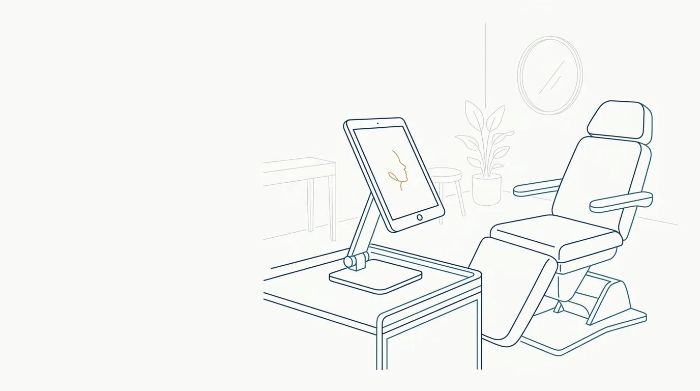 Showing the projection to the patient starts with the photoThe projection only convinces and aligns expectations if it starts from a standardized photo. See what to check in the photo before showing the slider.
 Side by Side or Slider Comparison: Which to Use in the VisitAdvantages and limits of side-by-side before and after versus a sliding comparison in the visit, and when to choose each format to show the patient.
 Smile photo in the facial assessment: what to recordWhat to record from the spontaneous smile in a facial assessment: teeth, lips, gums, symmetry, and folds, and how to standardize it to compare later.
 Speech video to assess lips and smileHow to record speech and smile to assess lip movement, incisor exposure and symmetry before and after lip procedures.
 Storing Patient Photos: Where to Keep Them SafelyFolder, cloud and encryption: how to organize and protect patients' clinical photos in the clinic, with restricted access and a review routine.
 Temple photo for documentation: angle and lightHow to photograph the temporal hollow with an oblique view and overhead light to document volume and the transition to the forehead and zygoma.
 Tense neck in the contour photo: effect on the double chinA forward chin, tense platysma and a stretched neck change the contour and double chin in the clinical photo. See how to standardize neck posture.
 To Edit or Not to Edit a Clinical Photo: Where the Line IsWhich adjustments to a clinical photo are acceptable (crop, alignment, exposure) and which become manipulation of the result, with a checklist.
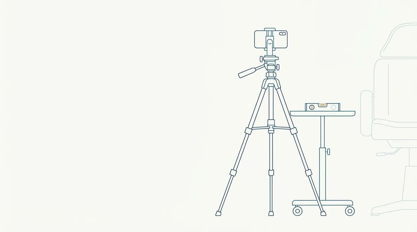 Tripod for clinical photos: phone height and levelWhy use a tripod for clinical photos, how to set the phone's height and level, what essentials to buy and how to repeat the position at every visit.
 Under-eye photo for documentation: light and angleHow to photograph under-eye circles and the tear trough with overhead light, a slightly raised chin and shadow control to document baseline and follow-up.
 Video to assess facial dynamics before botulinum toxinHow to record expressions to plan and follow up botulinum toxin: a movement script, slow-motion review, and comparison between sessions.
 When to take the follow-up photo after a procedureUsual time windows for the follow-up photo in facial harmonization, by product type, and how to record swelling, settling and results without being misled.
 White Balance in Clinical Photos: Lock It and StandardizeHow to use a gray card and manual mode to lock the phone's white balance and keep skin tone comparable between visits.
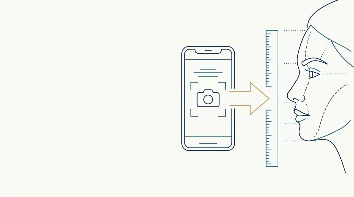 Wide-angle lenses distort the face in clinical photosA phone's wide-angle lens, used up close, widens the nose and forehead and shrinks the ears. How to avoid the distortion and which distance to use.
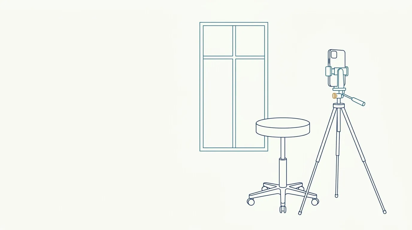 Window light as a light source in clinical photosSide window, time of day and daily variation: how to use natural light in clinical photos without harsh shadows or color differences between visits.