
Frankfurt plane in the clinical photo: positioning the head
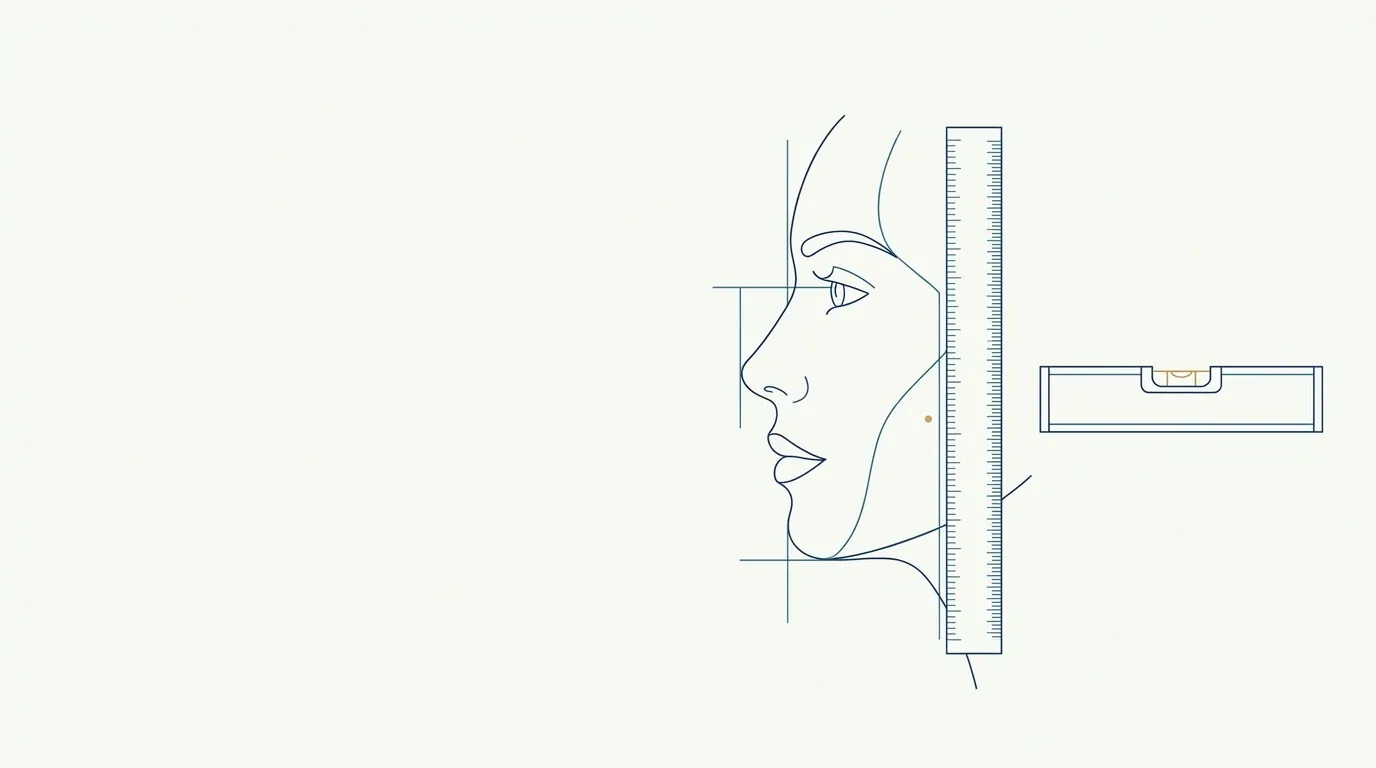
- Nerve
- Highlighted area
Quick facts
- Definition
- An imaginary line between the lower border of the orbit and the highest point of the external acoustic meatus.
- Goal in the photo
- That line parallel to the floor, with the head in natural position.
- How to check
- Compare the height of the outer corner of the eye with the tragus, or use a discreet level.
- Common mistake
- A raised or tucked chin, which tilts the plane and changes the contour and proportions.
When the patient raises the chin, the profile changes. When they tuck it, the double chin appears. These are effects of head rotation, and to control them facial analysis uses an anatomical reference line: the Frankfurt plane. It is the basis of almost every standardized clinical photo of the face.
You do not need to master cephalometry to use the concept in your routine. It is enough to know what it is, how to check it and when to adopt a different criterion.
What is the Frankfurt plane?
The Frankfurt plane is the line that joins the lowest point of the orbital rim, the orbitale, to the highest point of the external acoustic meatus, the porion. The name comes from the anthropology consensus in Frankfurt in the late nineteenth century, which adopted this line as the reference for head orientation.
In a person standing and looking at the horizon, the line is approximately parallel to the floor. That is why, in the standardized photo, this is the goal: a horizontal Frankfurt plane. The diagram on the page shows the points and the line, and the glossary entry on the Frankfurt plane covers the anatomical definition.
Why does it matter for HOF planning?
Because almost everything in the lower third depends on head rotation. Look at the effect of a small tilt.
| Head position | What the photo shows | Most affected area |
|---|---|---|
| Chin raised | Flatter profile, double chin less visible, nostrils exposed | Chin, nose, submental area |
| Chin tucked | Double chin and fold under the chin | Contour and submental area |
| Head tilted to one side | Apparent asymmetry | Eyes, mouth, jaw |
| Head turned | One side of the face looks larger | Cheekbone, jaw |
In planning for the chin, a raised chin in the photo leads to underestimating the need for projection. In assessing the under-eye area, looking up changes the shadow of the groove and the apparent volume of the lower eyelid.
How do you align the patient's head?
The simplest way does not require equipment. Follow the sequence.
- Seat the patient with an upright spine and loose shoulders.
- Ask them to look at a fixed point ahead, at eye level (see gaze at the horizon in the clinical photo).
- On the screen, find the outer corner of the eye and the tragus, the small cartilage bump in front of the ear canal.
- If the two points are at the same height, the plane is approximately horizontal.
- If the tragus is lower than the eye, the chin is raised. If it is higher, the chin is tucked.
- Adjust with a gentle cue, such as "lower your chin a little", and retake the shot.
The tragus is a surface reference, more practical than the porion. With ears of a different shape or covered by hair, checking becomes harder, hence the recommendation to tie the hair back, as in hair tied back in the clinical photo.
Frankfurt plane or natural head position?
There are two accepted choices in clinical photography practices. The first is the horizontal Frankfurt plane, an anatomical reference that is easy to check. The second is natural head position, which respects each person's spontaneous posture. The two usually coincide approximately in many patients, but in some the difference is noticeable.
For the HOF routine, what matters is choosing one criterion and keeping it. If the clinic uses Frankfurt, the follow-up photo uses it too. If it uses natural position, same thing. Mixing criteria between visits is the mistake, not the choice itself. The discussion on the gaze point is in gaze at the horizon.
How do you handle patients who cannot align?
Some people have neck limitations, scoliosis, marked posture or pain when holding the position. In those cases, the goal becomes repeatability, not perfection. Record the position the patient can sustain and repeat it in the following photos. Note the limitation on the sheet, so that another team member does not try to correct what cannot be corrected.
Avoid forcing the head with your hand without warning. If touching is necessary, ask permission, explain and use a light touch. The patient's comfort also improves the photo, because tension alters the expression.
The plane and the profile photo
The profile photo requires the same care, with the gaze at the horizon and the Frankfurt plane level. The profile is very sensitive to head rotation, and a few degrees alter the projection measurements of the chin and nose. When the practice takes profile photos, the extra care of using a wall reference and a level is worthwhile, covered in profile photo of the face at the clinic and in chin profile photo for planning.
Remember that Harmy uses only the frontal photo; it does not work with profile or 45-degree views. The app's projection shows the face from the front, with the marked areas.
And in Harmy?
The app shows marking points over the detected face and creates the projection from the frontal photo. If the head was rotated or the chin out of position, the simulation keeps that. For an honest projection, follow the same position criterion as the chart. The simulation is illustrative and at the discretion of the qualified professional, not a promise of results. The step-by-step is in standardized frontal photo. The overview of the topic is in standardized clinical photography.
Summary for the routine
Define the criterion (Frankfurt or natural position), check the corner of the eye and the tragus, ask for gentle adjustments and take more than one shot. Record the patient's limitations. With the head level, the profile, contour and periorbital region stop varying by chance.
Frequently asked questions
What is the Frankfurt plane?
It is a reference line in facial analysis and cephalometry that passes through the lower border of the orbit and the upper point of the external ear canal. It is used to orient the head.
Does the Frankfurt plane have to be parallel to the floor in every photo?
In standardized photos, yes, it is the most widely used reference. Natural head position may differ a little; what matters is defining the criterion and repeating it.
How can I align without a ruler or instrument?
Compare the height of the outer corner of the eye with the tragus, on the side of the ear. If they are on the same horizontal line, the plane is approximately level.
Are the Frankfurt plane and natural head position the same thing?
No. The plane is an anatomical line. Natural position is the spontaneous posture of someone looking into the distance. Each practice chooses one as its reference and sticks to it.
Harmy images are illustrative simulations generated by AI from the patient's photo to support the conversation between practitioner and patient. They are not a promise of results. Indication, technique, product and volume are solely the decision of the licensed practitioner. This content is for information only and does not replace a clinical assessment.