
Glabella Photo in Contraction to Document Botulinum Toxin
- Muscle
Quick facts
- Goal
- Record the glabella at rest and at maximum contraction, with the same cue at every visit.
- Cue
- Frown as if angry, holding for a few seconds.
- Poses
- Rest, maximum contraction and, if useful, light contraction, in the same light and distance.
- Follow-up
- Repeat the same poses to compare the response, without promising results.
In the glabella, what botulinum toxin treats is movement. That is why a single photo at rest tells little of the story: many lines between the eyebrows only appear when the patient frowns. To document the indication and, at follow-up, the response, the record needs at least two poses: rest and contraction. And it has to be done the same way at every visit.
This page describes the standard for photographing the glabella, the cues for the patient and the mistakes that spoil the comparison. The focus is documentation, not injection technique.
Which muscles show up in glabellar contraction?
The "angry" expression involves the corrugator muscle, which brings the eyebrows together, and the procerus, which pulls them down, along with other elements of the area. The visible result is vertical lines between the eyebrows and, sometimes, horizontal lines at the root of the nose. The contraction is part of dynamic wrinkles and is the reason for the indication of botulinum toxin. The clinical context of the area is in glabella and in toxin in the glabella.
Which poses should you photograph?
| Pose | Cue | What it shows |
|---|---|---|
| Rest | "Relax your face, look at the point on the wall." | Static lines and eyebrow position |
| Maximum contraction | "Frown as if you were angry, hard, and hold." | Extent and intensity of the dynamics |
| Light contraction (optional) | "Frown just a little." | Degrees of response |
| Raised eyebrows (optional) | "Raise your eyebrows." | Interaction with the frontalis; see photo of the forehead raising the eyebrows |
How do you standardize the cue?
The same instruction, in the same words, produces comparable contractions. A short script:
- Rest: "Look at the point and relax your forehead. I'm going to take the photo."
- Contraction: "Now frown as if you were angry, hard, and hold until I tell you."
- Release: "You can let go."
Note the cue in the medical record so another team member can repeat it. Avoid demonstrating the expression with your own face, because the patient tends to copy and exaggerate. The general expression script is in neutral expression in clinical photos and in photo of the face at rest and in motion.
Light, angle and distance
The glabella is an area with fine relief. The light needs to be soft and come from the front and slightly above, to show the furrow without creating harsh shadow. Avoid flash and strong raking light. Keep the Frankfurt plane horizontal, with the chin at its natural height, as in Frankfurt plane in clinical photos, and the lens at eye level (camera height).
Frame from the top of the head to the base of the nose at minimum, without bringing the lens so close that it distorts. A second, closer photo, zoomed by cropping and not by lens, helps to see detail.
How do you use the photo at follow-up?
At follow-up, repeat the same protocol of poses, light and distance. The comparison serves to see whether the contraction was maintained, decreased or whether there is asymmetry, without promising a result. The follow-up interval and the assessment are defined by the professional and vary with the product, and reading the image should come together with the clinical exam.
If the patient reports a drooping eyebrow or eyelid, the photo at rest and in elevation documents the situation. See also the complications in complications with botulinum toxin and the documentation page for toxin in dynamic photos for botulinum toxin.
What should you record in the medical record?
- date and time of the photos;
- poses taken and the cue used;
- device, distance and light;
- the patient's concern about the area;
- management defined by the professional;
- consent for use of the image, per consent and LGPD in photos.
And in Harmy?
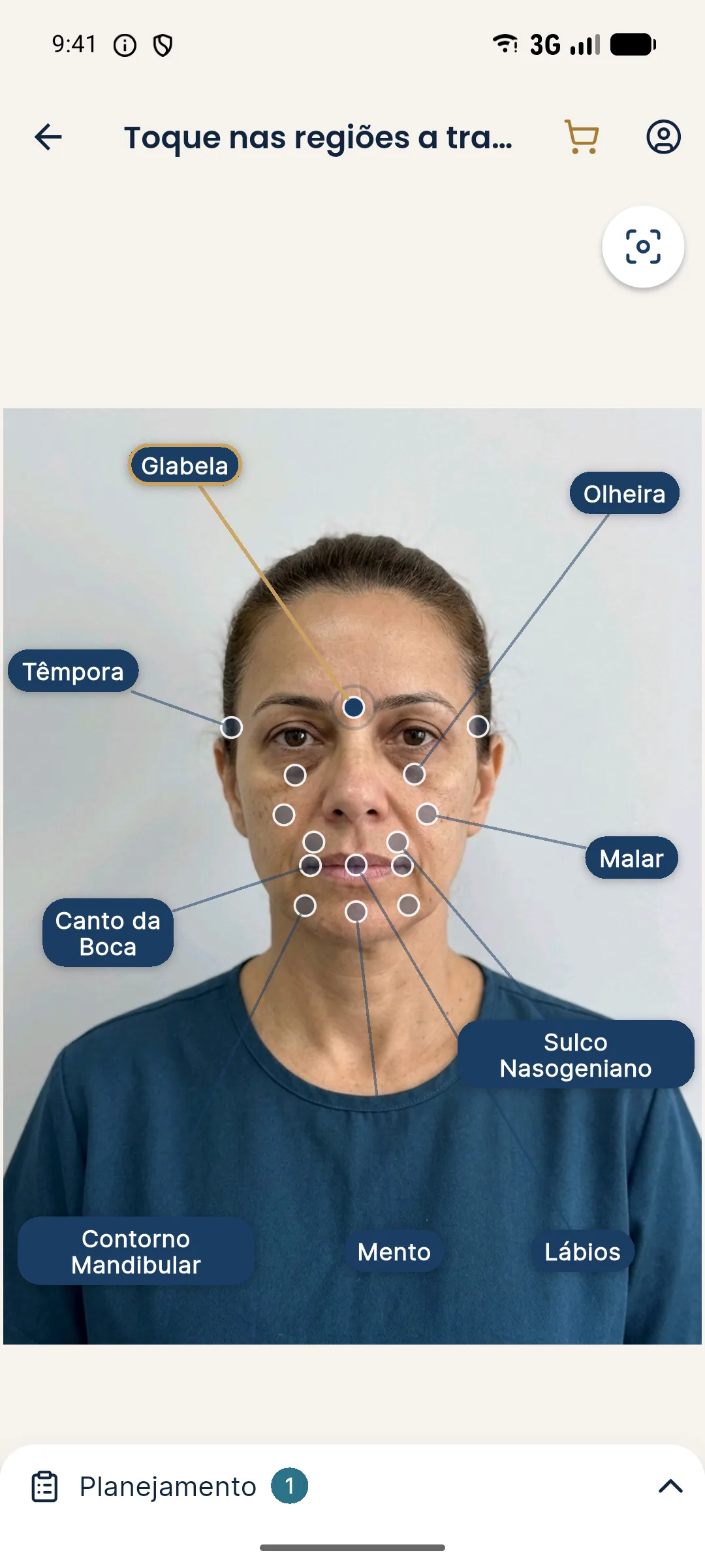
The app uses a frontal photo at rest and lets you mark the "Glabella" point with the symbolic volume. The illustrative simulation shows a static projection of how the area may look, from the original photo, and does not represent movement or the effect of the toxin in contraction. The contraction photo is a separate documentation record.
Common mistakes in the contraction photo
The first is asking for the contraction without saying when to release, which makes the patient hold the tension and tire the muscle. The second is photographing the contraction before rest, leaving the glabella marked. The third is varying the strength of the request from one visit to another: "frown a little" at baseline and "frown hard" at follow-up produce comparisons that do not hold up. The fourth is allowing head tilt, which changes the angle of the lines. A written script repeated by the team solves most of these problems.
Summary
Two poses, the same cue, soft light and complete notes. With this standard, the glabella photo documents the patient's dynamics and lets you compare the response at follow-up honestly.
Frequently asked questions
Why photograph the glabella in contraction?
Because the contraction shows what the toxin is meant to act on and documents the effect at follow-up. At rest, many lines look subtle, and only contraction reveals the dynamics.
What cue should I use for glabella contraction?
Ask the patient to frown as if they were angry, holding the expression for a few seconds while you shoot. Always use the same words.
How many glabella photos should I take?
Generally two poses, rest and maximum contraction, with a short burst of each. Some routines add a light contraction to compare degrees.
Can I compare the contraction photo with Harmy's simulation?
Harmy's simulation uses the photo at rest and does not represent the effect of the toxin in contraction. The contraction photo is a documentation record, kept separate.
Harmy images are illustrative simulations generated by AI from the patient's photo to support the conversation between practitioner and patient. They are not a promise of results. Indication, technique, product and volume are solely the decision of the licensed practitioner. This content is for information only and does not replace a clinical assessment.