
Ideal Malar Projection: How to Set the Goal in the Plan
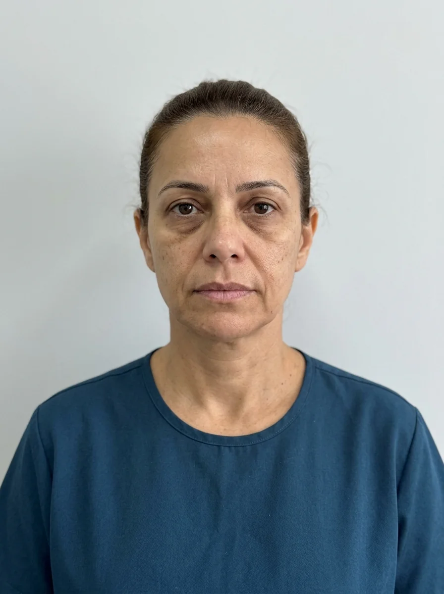
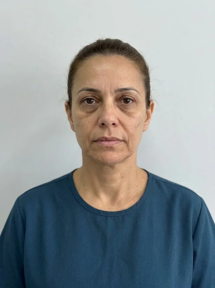 BeforeSimulation
- Highlighted area
Quick facts
- Topic
- Deciding how much and where to project the cheekbone before choosing volume and intensity
- References
- Hinderer lines, point of greatest projection, projection vector and the triangle of youth
- Decision
- Vertical and lateral projection are different goals, with distinct effects on the gaze and the midface
- Caution
- Projecting beyond what bone and skin can support may look artificial and alter the patient's identity
The malar region is the area of the cheekbones, one of the pillars of the midface. When it is well supported, the face looks more open, and the shadows under the eye and beside the nose tend to soften. When projection is lacking, the midface looks flat and tired. The planning question is: what is the projection goal, and where should it be?
This page organizes the reference points, tells anterior from lateral projection and sets out the limits of what volume can do. It does not cover injection technique.
What is the logic of a projection goal?
A goal is a target that combines three things: what the bone allows, what the skin supports and what the patient recognizes as their identity. If the goal ignores any one of them, the result loses naturalness. A heavily projected cheekbone over thin, fragile skin, for example, can end up looking like artificial volume, which is described in pillow face and how planning avoids it.
Setting the goal starts with assessing the skeleton, before the soft tissue. See facial skeleton before injecting and the anatomy of the zygomatic bone and the zygomatic arch.
Which reference points help locate the cheekbone?
The most-used references are lines and triangles that help you read the area. They serve as a visual guide, not a rule.
| Reference | What it shows | How to use it |
|---|---|---|
| Hinderer lines | Point of greatest malar projection | Upper lateral quadrant of the crossing between the mouth corner-lateral canthus line and the alar base-tragus line |
| Projection vector | Direction in which the volume stands out | Forward, lateral or combined |
| Triangle of youth | Midface volume in a young face | Wide base at the cheekbones and apex at the chin; with age, it tends to invert |
| Medial malar compartment | Superficial fat of the cheek | Where volume tends to descend with age |
The use of Hinderer lines in assessment is described on its own page, Hinderer lines in malar assessment. One caveat: the Hinderer scheme was described in a specific context, and adapting it to other populations requires care.
Anterior or lateral projection: which to choose?
They are two different goals, with distinct effects.
- Anterior projection (forward): increases volume on the frontal face and in profile, softening the flat look of the midface. It is the projection most associated with rejuvenation and with support of the infraorbital area.
- Lateral projection (sideways): increases the width of the face at the level of the zygoma, which changes the shape of the face. In faces that are already wide, it can worsen the sense of width; in narrow faces, it can balance.
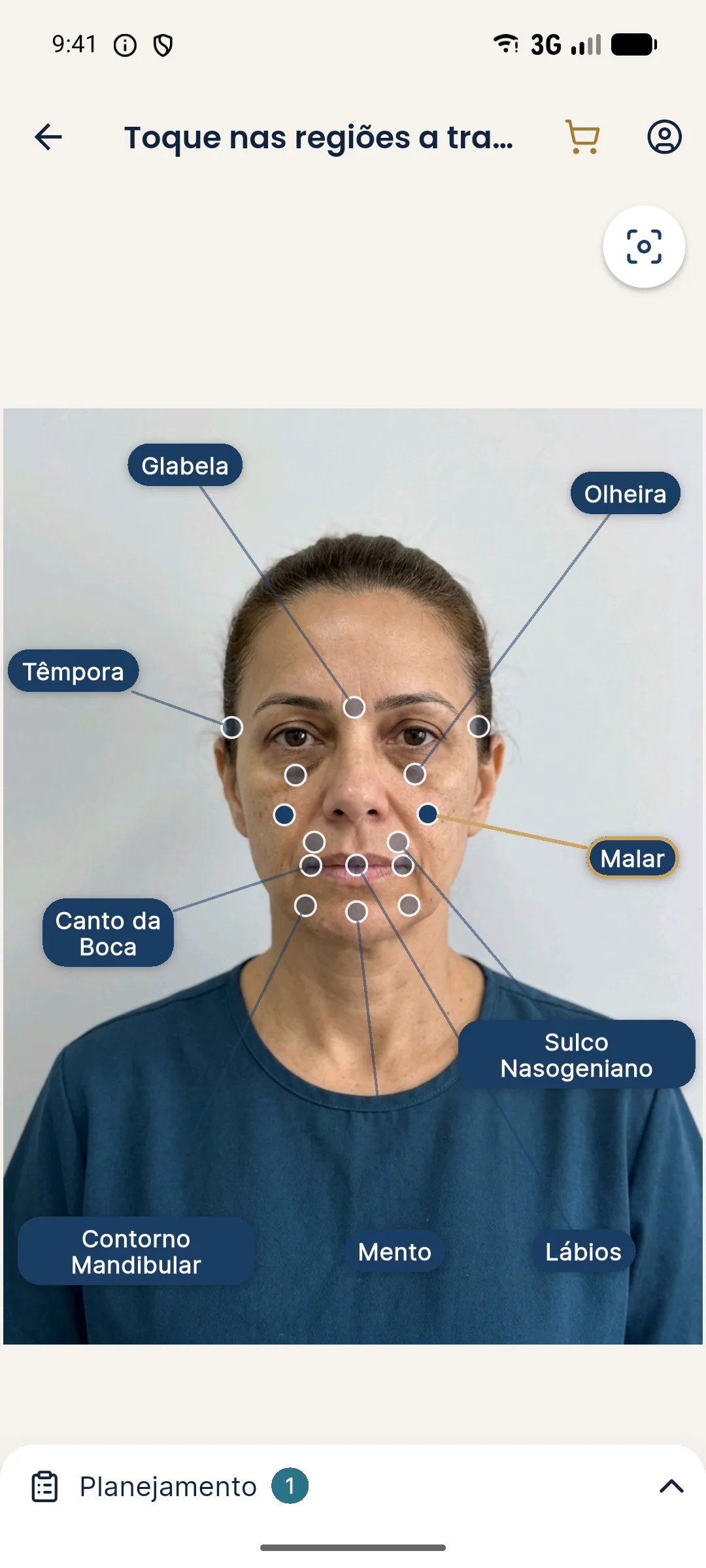
The choice depends on face shape. The reasoning by shape is in planning a wide face, long face, oval face and round face. In Harmy, the projection is made on the frontal photo, and lateral projection shows up there more than anterior.
How do age and sex shape the goal?
Over time, the cheekbone loses volume and support, and fat descends. This changes the goal: in young patients, projection is usually a decision about shape; in mature patients, about support. The approach by age group is in facial analysis at 40, at 50 and at 60 or older.
Between the sexes, the described trend is a more projected, higher cheekbone in women and a flatter, wider one in men. This is a textbook trend, not a rule, and changing how the face reads in terms of gender without the patient's intent is a planning error. See volume by sex in planning.
What can volume do, and what can it not do?
Dermal filler adds volume. It does not add bone. When the deficit is skeletal, volume has a ceiling, and going past it leads to a puffy effect instead of projection. When the deficit is in soft tissue, volume tends to work better. The difference is covered in when bone is the problem, not volume, and malar hypoplasia has its own entry.
The texts malar filler and zygomatic filler detail what is done in each case. As with any area of the face, risks include swelling, bruising, asymmetry and, rarely, vascular complications, and hyaluronidase must be on hand when hyaluronic acid is used.
How does the cheekbone relate to the rest of the midface?
The cheekbone supports the tear trough, the nasolabial fold and the transition to the cheek and jaw. Changing the cheekbone often improves these areas indirectly, and this is part of the reasoning in cheekbone and tear trough: combining them in the plan, nasolabial fold and cheekbone: combining them in the plan and cheekbone and jaw: combining them in the plan. The whole picture is in complete midface in the plan.
This interdependence is useful and dangerous at the same time. It is useful because it lets you solve more with less. It is dangerous because it gives the impression that the cheekbone fixes everything, which can lead to excess volume. See the warning in when total volume becomes excess.
How to talk to the patient about the goal?
Show the starting image, point out what the area supports and make clear that the cheekbone is part of a whole. Patients often ask for "higher cheekbones" without knowing that this changes the width of the face. Explain the difference and show the options. The wording is in explaining the cheekbone to the patient.
Frequently asked questions
What is the ideal malar projection?
There is no single value. Reference lines help locate the point of greatest projection, but the goal depends on bone, skin, sex, age and what the patient recognizes as their own.
Does projecting the cheekbone fix tear troughs and the nasolabial fold?
It can contribute, because the cheekbone supports the midface. Even so, the tear trough and the fold have their own causes, and each concern deserves an individual assessment.
What is the difference between projecting forward and projecting sideways?
Anterior projection changes volume on the frontal face and in profile; lateral projection increases the width of the face at the level of the zygoma. Each changes how the whole face reads in a different way.
Does Harmy simulate malar projection?
The Cheekbone marking lets you choose ml and intensity, and the projection shows an approximation on the frontal photo. Anteroposterior projection remains a clinical assessment.
Harmy images are illustrative simulations generated by AI from the patient's photo to support the conversation between practitioner and patient. They are not a promise of results. Indication, technique, product and volume are solely the decision of the licensed practitioner. This content is for information only and does not replace a clinical assessment.