
Hinderer's lines in malar assessment
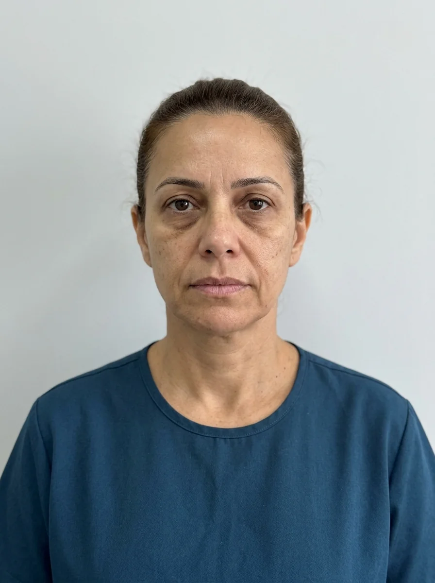
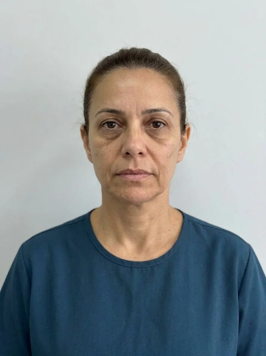 BeforeSimulation
- Highlighted area
Quick facts
- What they are
- Reference lines used to locate ideal cheekbone projection relative to fixed points on the face
- Use
- Reading guide to decide where malar projection seems to be lacking or excessive
- Limit
- A classic proportion reference: it varies between people and populations and is not a target
- Warning
- A well-projected cheekbone does not depend on volume alone: bone, fat, skin, and ligaments all count
The question "where should the highest point of the cheek be?" is an old one in facial aesthetics. Many references have been proposed over time to help answer it: lines, triangles, angles. Hinderer's lines are among the best known in the malar assessment literature, and they show up often in harmonization courses.
This page presents what these lines are, where they came from, how to use them as a reading guide, and above all what their limits are. The goal is to get the benefit without falling into the trap of turning a classic reference into an injection rule. It is not a protocol: the decision belongs to the qualified professional, after assessment.
What are Hinderer's lines?
They are reference lines drawn over the face that help locate the area of greatest cheekbone projection. In Hinderer's description (a plastic surgeon, in a 1975 paper on malar implants), two lines are drawn: one from the mouth corner to the lateral canthus of the eye, and another from the base of the nasal ala to the tragus. Their crossing divides the cheek into four quadrants, and the area of greatest malar projection is expected in the upper lateral quadrant.
The description is offered as a reading reference: there is no universal exact measurement, and later courses and authors make small adaptations. The proposal was born in the surgical planning of malar implants and came to be used for the aesthetic reading of the midface as well. The bony structure involved is covered in zygomatic bone and zygomatic arch.
What are they used for in practice?
The most honest use of the lines is in reading the photo and the live exam, as support.
- Locating the projection. Is the highest point of the cheek higher, lower, more inward, or more outward than expected?
- Noticing midface descent. When volume descends with age, the projection point descends with it.
- Organizing the conversation. Showing the patient, on their own photo, where volume seems to be lacking.
- Comparing before and after. Checking whether the projection stayed balanced.
| Finding when drawing the lines | Possible reading |
|---|---|
| Projection in the expected area | Balanced midface |
| Lower and more medial projection | Possible tissue descent |
| Little projection overall | Possible volume loss or a less projected structure |
| Very lateral projection | Wide bony structure or lateral excess |
This table is a thinking aid, not a diagnosis. The midface is covered in detail in complete midface in the plan, and malar projection in ideal malar projection.
What are the limits of the method?
The limits are significant and should be stated clearly.
- Variation between people. Bone structure, fat, and skin vary, and there is no "right" point that holds for everyone.
- Variation between populations. The shape of the zygomatic bone and average projection differ between groups, and a reference built on one group may not suit another. See facial proportions vary between populations.
- Dependence on the photo. The tracing changes with angle, distance, and expression.
- Simplification. It reduces a three-dimensional structure to two lines.
- It ignores the cause. Lack of projection may be bony, fat-related, skin-related, or due to tissue descent, and the lines do not tell these apart.
How do you use them carefully in malar assessment?
A short, prudent routine:
The photo and the reading need standardization, as described in standardized clinical photography and oblique (three-quarter) facial analysis. For the full set of reference lines on the frontal photo, see reference lines on the frontal photo.
And the cause of the lack of projection?
The same impression of a "flat cheekbone" can have different origins, and management changes with the cause.
| Cause | How to think about it |
|---|---|
| Less projected bony structure | Volume may help, within limits; assess expectations |
| Loss of fat volume | Volume replacement may be considered |
| Tissue descent | May call for support before volume |
| Thin, lax skin | Skin quality may be the priority |
| Combination | Plan in stages |
The distinction between sagging and volume loss appears in ptosis or volume loss: which to treat, and the fat compartments in midface fat compartments in the plan. The youth triangle is another reference that helps explain the change in the midface.
What precautions does the area require?
The midface has important structures: the transverse facial artery, branches of the facial nerve, the infraorbital nerve, and proximity to the periorbital region. Possible complications include swelling, bruising, asymmetry, irregularities, and, rarely, vascular complications. With hyaluronic acid filler, hyaluronidase must be available. See periorbital anatomy for injectables and safety and complications: overview.
The decision on technique, plan, and volume rests with the qualified professional, according to the rules of their professional council.
How do you explain it to the patient?
Patients usually grasp the idea of a "map" well. Show them on their photo where the lines cross and talk about what that suggests, without promising anything.
The conversation about the cheekbone appears in explaining the cheekbone to the patient.
How does Harmy fit into malar assessment?
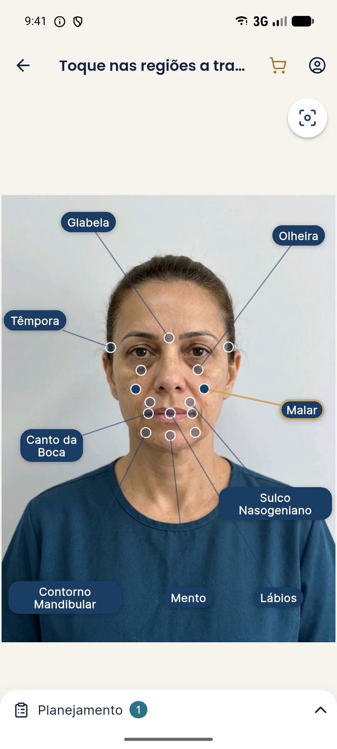
Use the lines in the exam and on the photo, and the app to illustrate volume in the conversation. The steps are in marking the malar area in Harmy.
Quiz: Hinderer's lines
Summary
- Hinderer's lines are classic references for reading cheekbone projection.
- They serve to locate projection and organize the conversation, not to define where to inject.
- They vary between people and populations, and depend on the photo.
- Identify the cause of the lack of projection before deciding.
- The area carries vascular and anatomical risk: keep hyaluronidase available.
- Harmy marks the Malar point without drawing reference lines.
Frequently asked questions
What are Hinderer's lines?
They are two lines drawn on the face: one from the mouth corner to the lateral canthus of the eye, and another from the base of the nasal ala to the tragus. Their crossing divides the area into quadrants, and the greatest malar projection is usually read in the upper lateral quadrant. They serve as a reading guide.
Do Hinderer's lines define where to place the filler?
No. They guide the reading of projection, but the plan depends on the live exam, individual anatomy, and the decision of the qualified professional. They are not an injection protocol.
Does every face need to fit the lines?
No. Variation between people and populations is large, and the goal of the plan is balance with the patient's identity, not fitting a ruler.
Does the app use Hinderer's lines?
No. Harmy does not draw reference lines. The Malar point lets you mark the area and choose ml and intensity, and the projection is an illustration.
Harmy images are illustrative simulations generated by AI from the patient's photo to support the conversation between practitioner and patient. They are not a promise of results. Indication, technique, product and volume are solely the decision of the licensed practitioner. This content is for information only and does not replace a clinical assessment.