
Midface Fat Compartments in the Facial Harmonization Plan
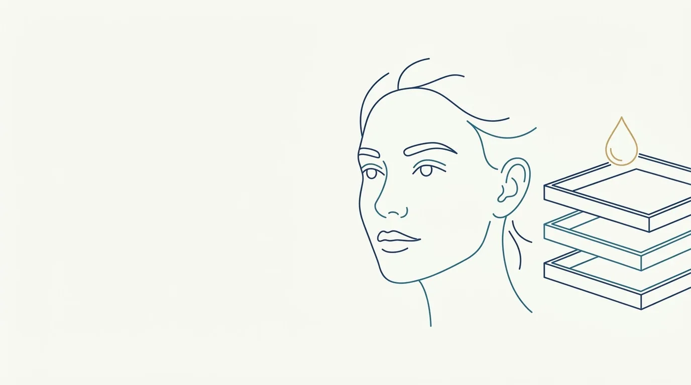
- Highlighted area
Quick facts
- Topic
- Midface fat compartments
- Division
- Superficial and deep
- Use in the plan
- Understand where volume was lost
- Caution
- Vascular area, technique is not covered on this page
When the patient says "my face is sagging" or "my under-eye got worse", the cause is almost never where they point. In the midface, a tired look, a deep fold and a flattened cheekbone are often the result of volume loss in fat compartments and of weakening of the ligaments that hold them. Reading these compartments is a way to plan with logic, instead of chasing the symptom.
What are fat compartments?
Facial fat is not a continuous layer. It is organized into compartments, separated by fibrous septa and ligaments. Some compartments are superficial, closer to the skin, and others are deep, next to the supporting structures. Each ages at its own pace: some lose volume, others descend, and the result is the change in contour we recognize as aging.
Among those most often cited in the midface are the nasolabial compartment, the deep medial cheek fat, the SOOF (sub-orbicularis oculi fat) and the other fat pads. The ligaments and spaces such as the prezygomatic space complete the map. The overview by layers is in facial aging by layers.
How does this change how you read the face?
Instead of asking "where is the fold?", you ask "what supported this area and lost volume?". This reorganizes the reasoning:
| Patient's concern | Cause question | Area to consider |
|---|---|---|
| Deep nasolabial fold | Was there loss of support above, in the cheekbone? | Cheekbone before the fold, when indicated |
| Pronounced tear trough | Has the lid-cheek transition lost volume? | Tear trough, with caution, and cheekbone |
| Tired face | Has the midface lost projection as a whole? | Cheekbone and zygomatic area |
| "Sagging" cheek | Have the compartments descended or shrunk? | Joint assessment of support |
The discussion about treating the cause is in treating the fold or the cause of the fold and the layering logic in structure before detail.
Superficial or deep compartment: what is the role of each?
Deep compartments support the midface, and their loss tends to show up as flattening and descent of the whole. Superficial ones shape the skin and the visible contour, and their change can form folds and bags. In practice, the plan usually considers support before surface. This order appears in structure before detail and in complete midface in the plan.
And when bone is the problem?
Not every loss of contour is fat. Bone resorption also changes the midface, and filling soft tissue over reduced bone support may not deliver what the patient expects. See also when bone is the problem, not volume and facial skeleton before injecting.
How do you use this reading in the plan?
The patient's old photo is useful for identifying what has changed. See patient's old photo as a reference. The volume decision comes afterward, in ml per area: ranges in planning.
Where does Harmy come in?
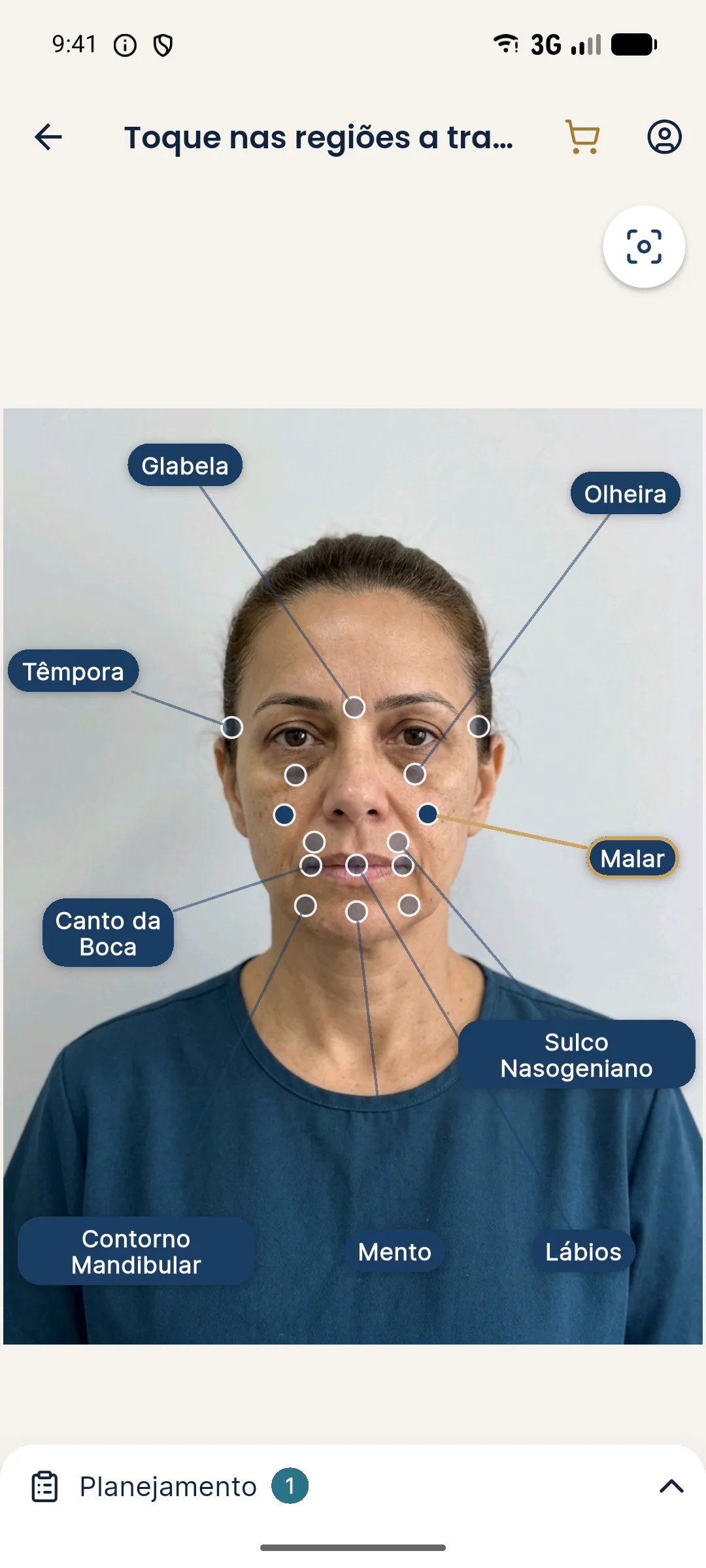
Harmy marks the Tear Trough, Cheekbone and Nasolabial Fold points on the frontal photo, with ml per area and intensity. The projection shows the change only in the marked areas. It does not identify compartments or estimate volume loss: the anatomical reading is the professional's. The most interesting use is comparing two scenarios, for example fold only versus cheekbone with fold, to show the patient why the order matters.
See also cheekbone and tear trough: combining in the plan and nasolabial fold and cheekbone: combining in the plan.
How does aging affect each compartment?
Without going into technique, the general pattern helps in reading the face. Over time, some superficial compartments lose volume and others descend, while the deep compartments shrink and stop projecting the face. The retaining ligaments, which anchor the skin to the bone, stay relatively stable and create "anchor points" where the skin folds, forming creases. That is why the nasolabial fold, the palpebromalar groove and the mid-cheek groove tend to appear along ligament-related lines.
Two practical consequences for the plan:
- The fold is, in part, a contrast effect. When the neighboring compartment loses volume, the crease at the ligament becomes more evident. Treating the compartment may change how the fold reads.
- Not every fold responds to the same approach. What fits each case depends on anatomy, skin, age and concern.
For further reading, see mid-cheek groove, palpebromalar groove and retaining ligaments.
One final caution: reading compartments is a model, and models simplify. Individual variation, previous surgeries, earlier use of fillers and rapid weight loss (see planning HOF in people who lose weight fast) alter the expected pattern. When in doubt, the safest option is usually a conservative first step followed by reassessment.
Summary
Reading the midface fat compartments transforms the plan: from the symptom to the cause. Observe, compare, prioritize support, start conservatively and reassess. Product, volume and technique are at the discretion of the licensed professional, after assessment, according to the rules of their professional board in Brazil.
Frequently asked questions
What are the fat compartments of the face?
They are areas bounded by septa and ligaments that store fat in superficial and deep layers. Each compartment ages differently, and reading them helps you understand the contour of the face.
Why does this matter in planning?
Because volume loss is not uniform. Understanding which compartment has shrunk helps you choose the area to treat, instead of filling where the patient sees the symptom.
Is the deep compartment more important than the superficial one?
It is not a question of importance. The deep ones provide support and the superficial ones influence the contour of the skin. The plan usually considers the relationship between the two.
Does Harmy identify fat compartments?
No. Harmy marks areas such as Cheekbone, Tear Trough and Nasolabial Fold on the frontal photo and generates an illustrative projection. Reading the compartments is the professional's job.
Harmy images are illustrative simulations generated by AI from the patient's photo to support the conversation between practitioner and patient. They are not a promise of results. Indication, technique, product and volume are solely the decision of the licensed practitioner. This content is for information only and does not replace a clinical assessment.