
Flat cheekbones: assessing midface projection
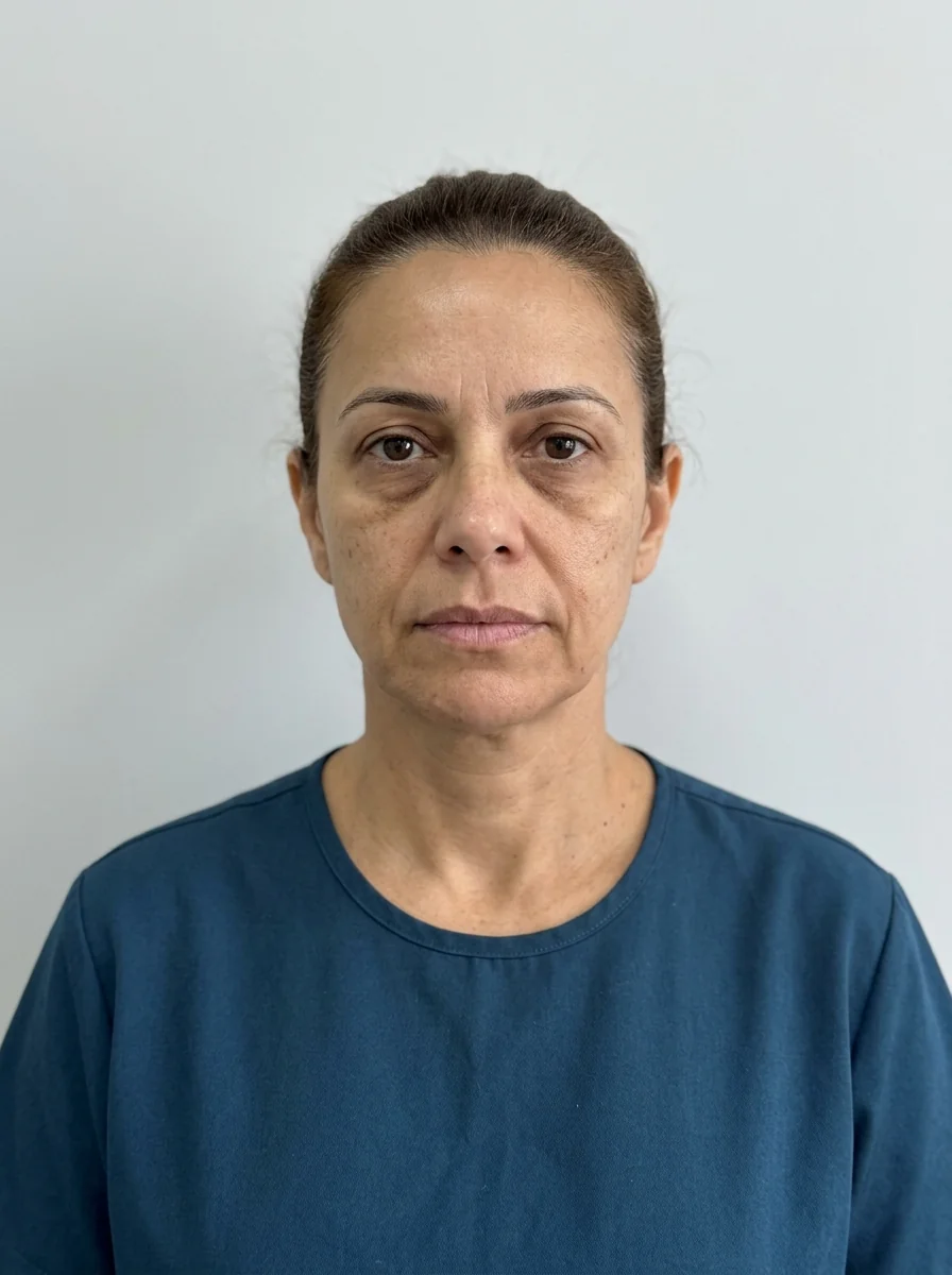
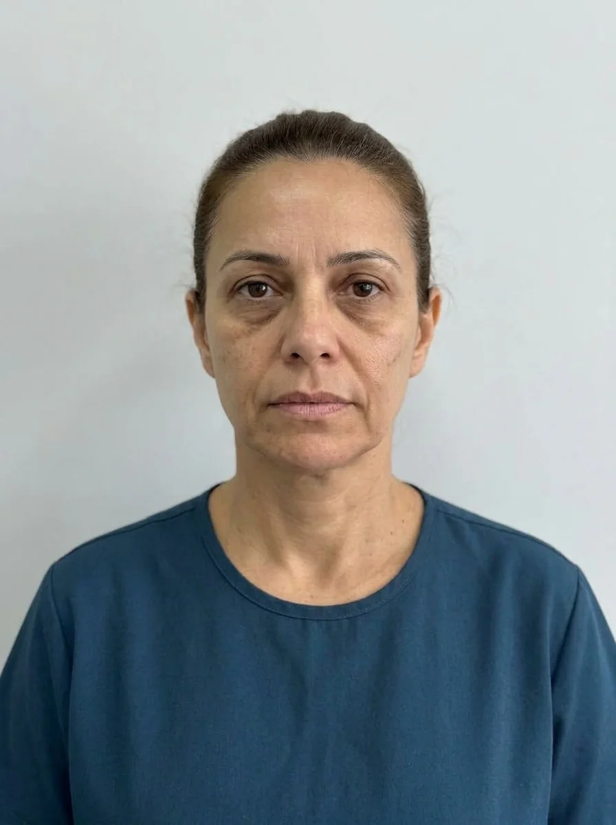 BeforeSimulation
- Nerve
- Highlighted area
Quick facts
- Concern
- The face looks flat from the front, with no projection of the cheekbone
- Method
- Assess from the front, three-quarter and profile views, separating bone from fat
- Structures
- Zygomatic bone, prezygomatic space, SOOF and malar fat
- Caution
- Too much projection widens the face and weighs down the midface
Flat cheekbones are a concern that shows up in two very different profiles: the young person whose bone has always been poorly projected, and the older person who has lost fat and support. In both, the face looks "flat from the front," with little light and shadow over the cheekbones, and the eyes take on a tired look.
This page organizes the assessment of the midface. It connects with low cheekbones, high cheekbones and hollow cheeks and profile without midface projection.
What makes the cheekbone look flat?
Projection of the midface depends on a bony base, the zygomatic bone, and on the layers of fat that cover it, such as the prezygomatic space and the SOOF. Flat cheekbones often combine:
| Component | Change | Clue in the assessment |
|---|---|---|
| Poorly projected zygomatic bone | Flat structural base | Flat profile since youth |
| Little malar fat | Volume loss | Angular face, pronounced under-eye hollow |
| Fat descent | Volume shifts downward | Deep nasolabial fold |
| Thin or lax skin | Poorly defined contour | Flat light, altered texture |
The relationship with the under-eye area matters: the palpebromalar groove tends to become more visible when the cheekbone loses support. Treatment of the under-eye area often starts with the cheekbone, as in cheekbone and under-eye: combining in the plan.
How to assess projection
Avoid assessing only from the front. Flat cheekbones are usually more evident in the three-quarter view, where you can see the transition from the zygoma to the cheek.
What the plan can consider
The plan starts from the base. At the discretion of the licensed professional:
- Zygomatic projection to restore structure, in a restrained amount. See zygomatic filler.
- Malar volume to soften the contour and the transition to the under-eye area. See cheekbone filler.
- Skin quality and gradual support, with collagen biostimulator in selected cases.
- Structure before detail: address the groove and the under-eye area only after providing support.
In a wide face, lateral projection calls for caution, and anterior projection (further forward) may be the better choice, because it gives light to the cheekbone without widening. The decision rests on each patient's assessment.
Risks
Swelling, bruising, asymmetry, nodules and irregularities are part of the picture. The area receives branches of the transverse facial artery and of the infraorbital nerve, and vascular complications are rare but serious. Hyaluronidase should be available when hyaluronic acid is used. Near the lower eyelid, thin skin favors the Tyndall effect and persistent swelling.
How to show it in Harmy
In Harmy, the Cheekbone point simulates support of the cheek in the front photo. It is the ideal tool for talking with skeptical patients: a projected cheekbone at Subtle intensity often already shows the difference in light. The "Maintenance" shortcut proposes the cheekbone with 1 ml at Subtle intensity, and "Full harmonization" proposes the cheekbone with 2 ml at Moderate, only as starting points.
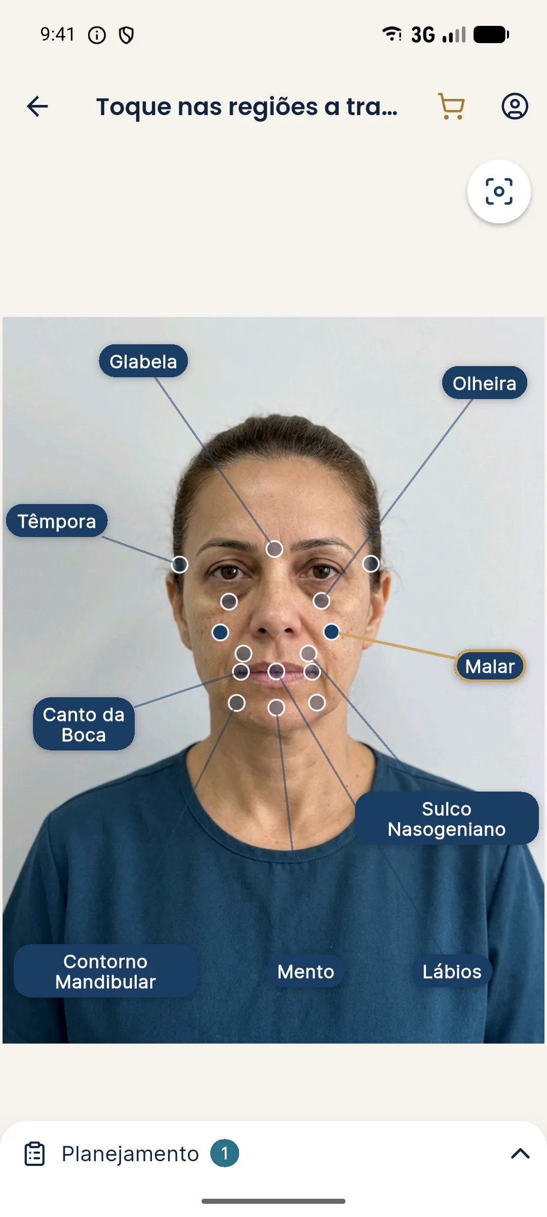
Make two projections, Subtle and Moderate, and use presentation mode to turn the phone around. In a bilateral area, the ml is the total split between the two sides.
Common mistakes with flat cheekbones
The first mistake is planning while looking only from the front. Midface projection is assessed in three-quarter and profile views, and frontal light flattens any face. The second is using the same recipe for everyone: the flat cheekbone of a young person, caused by bone, does not respond like that of an older person, caused by fat loss.
Another mistake is projecting too much to the side, widening a face that was already wide. In many cases, anterior projection, which gives light to the cheekbone without increasing width, is more suitable. It is also a mistake to ignore old fillers, which may be changing the shape of the midface without the patient remembering.
Finally, avoid promising a "younger" face. A well-worked cheekbone can provide support and light, but the patient should understand that this is about proportion and that results vary.
Follow-up and evaluating the result
Schedule the follow-up to reassess projection, symmetry and the relationship with the under-eye area, always with the same light and position. Record the assessment from three angles, the plan and what was agreed as the next stage. If the patient wants more volume, discuss the risk of an artificial look and suggest waiting, because the initial swelling can distort perception. Document the consent, which should mention asymmetry, nodules, persistent swelling and vascular risk. In patients who are concerned about their smile, review the dynamics before any add-on. Patience with the cheekbone usually pays off in a more natural and longer-lasting result.
Patient profiles and strategies
In young patients with a poorly projected bone, the conversation centers on structure and proportion, and the approach can focus more on the bone. In patients with fat loss, the focus is on support and the transition to the under-eye area and the fold.
Patients with thin skin and a pronounced under-eye hollow benefit from plans that start with the midface, but they require careful technique because of the risk of persistent swelling and Tyndall. Patients with a wide face call for anterior rather than lateral projection.
Before any decision, show the patient photos with side and frontal light, so that they understand why light changes the perception of the cheekbone. This helps align expectations and choose the most conservative path.
Wrap-up
Record the photos with side light, the perceived bony base, the fat layer and the staged plan. For related cases, see sunken cheeks and face that looks flat from the front. Volumes and techniques are at the discretion of the licensed professional, in accordance with the rules of their professional council in Brazil.
Frequently asked questions
Are flat cheekbones caused by a lack of bone or of fat?
It can be both. The zygomatic bone sets the base, and the malar fat gives the contour. Palpation, the profile view and three-quarter photos help tell them apart.
Does filler fix flat cheekbones?
It can improve projection and support of the midface, at the discretion of the licensed professional. The volume should be restrained and the patient's identity respected.
Is there a risk of making the face too wide?
Yes, especially when volume is placed laterally in a face that is already wide. Measuring beforehand and simulating lower intensities helps prevent it.
Is a biostimulator an option for flat cheekbones?
It may be considered in suitable cases, at the discretion of the licensed professional, for gradual quality and support, without replacing the assessment of the underlying structure.
Harmy images are illustrative simulations generated by AI from the patient's photo to support the conversation between practitioner and patient. They are not a promise of results. Indication, technique, product and volume are solely the decision of the licensed practitioner. This content is for information only and does not replace a clinical assessment.