
Low cheekbone apple: assessing the malar point
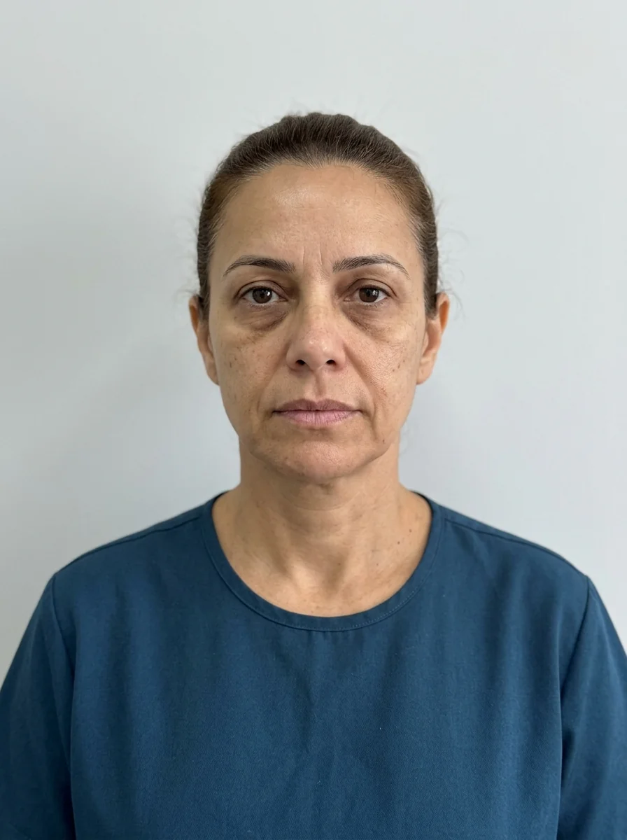
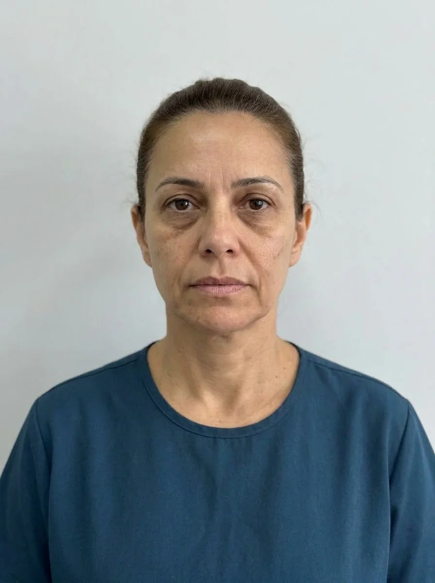 BeforeSimulation
- Highlighted area
Quick facts
- Concern
- The apple of the cheek looks low, flat or displaced toward the mouth
- Method
- Locate the point of greatest projection in three-quarter and profile views and compare it with the old photo
- Structures
- Zygomatic bone, malar fat, SOOF and ligaments
- Caution
- Raising the volume too much can widen the face or create a ball-like look
When the patient describes that "the cheek dropped," they are not always talking about laxity. Sometimes what changed was the point of greatest malar projection, the "apple," which migrated downward or faded. Visually repositioning that point is one of the effects most associated with midface rejuvenation.
This page shows how to locate the point, what may have dropped and how to think through the plan. See also sagging cheeks, flat cheekbone and midface ptosis.
Where is the malar point?
The malar point is the area of greatest projection of the zygomatic bone covered by fat and skin. In a young face, it sits relatively high and lateral, with a smooth transition to the temple and to the nasolabial fold. References used in practice include the line between the corner of the mouth and the outer corner of the eye, and the relationship with the midface seen from the side.
| Situation | What is observed | Possible component |
|---|---|---|
| High, defined apple | Lateral projection, full cheek | Youthful pattern |
| Faded apple | Flat face from the front | Loss of fat and bone |
| Low apple | Volume concentrated near the mouth | Fat descent (SOOF and malar) |
| Wide, low apple | Poor definition, wide face | Excess soft tissue, laxity |
The volume descends because the zygomatic ligament gives way and the fat of the SOOF and the malar area pools lower. This reinforces the nasolabial fold and creates a "heavy" face look.
How to locate and compare
Assessing the smile is part of the plan. The reasoning is in cheek without volume when smiling. The Hinderer lines are a classic reference for the position of the point.
What harmonization can consider
At the discretion of the licensed professional, the plan may combine:
- Zygomatic bone projection, with volume in restrained amounts, to create the perception of a higher apple. See zygomatic filler.
- Midface support, with cheekbone filler, in stages.
- Skin quality and collagen stimulation, in selected cases.
- Nasolabial fold, treated after support, because it often improves on its own.
A frequent mistake is filling the fold before giving support to the cheekbone, and the result is a heavier face. The logic of "structure before detail" appears in structure before detail in the harmonization plan.
Risks
Swelling, bruising, asymmetry, nodules and irregularities; rare and serious vascular complications, because the area has branches of the transverse facial artery and vessels close to the eye. The infraorbital nerve calls for care. Hyaluronidase must be available when hyaluronic acid is used. Proximity to the tear troughs requires attention to swelling and the Tyndall effect.
How to show it in Harmy
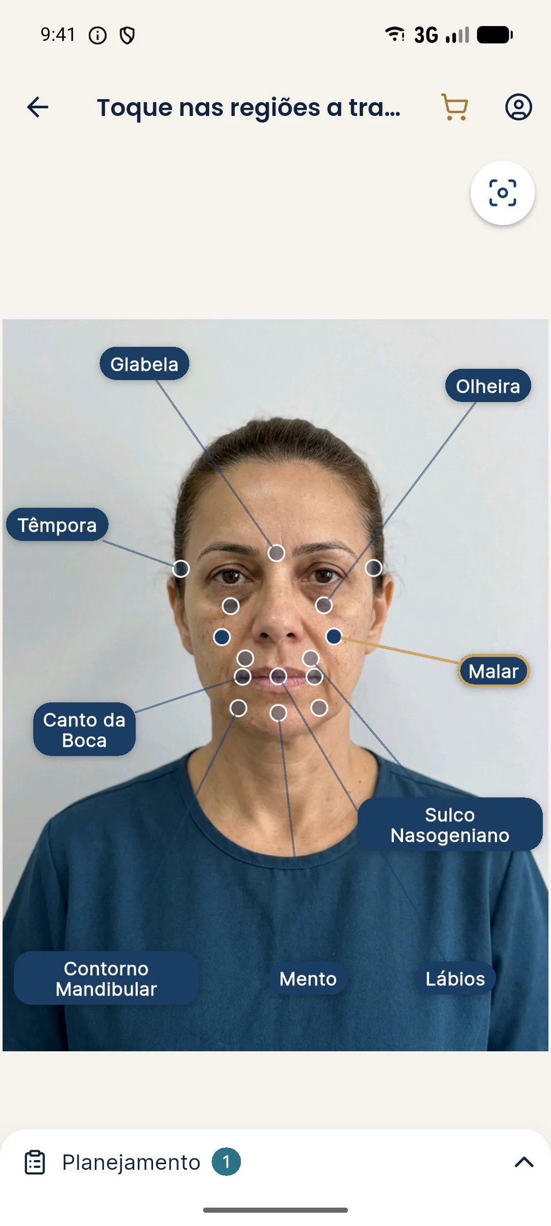
The Cheekbone point in Harmy simulates support of the apple on the frontal photo. In the frontal view, the app illustrates lateral volume, but does not measure the height of the malar point. The "Maintenance" shortcut suggests cheekbone at 1 ml with Subtle intensity, and "Full harmonization" suggests cheekbone at 2 ml with Moderate, which can serve as a conversation starting point, with no recommended dose.
Common mistakes when repositioning the apple
The most frequent mistake is concentrating volume at the wrong point. Placing product too low, near the mouth, reinforces the descent and accentuates the nasolabial fold; too lateral, it widens the face. Locating the high point requires a three-quarter photo and comparison with the patient's own reference, not with a generic image.
The second mistake is treating the fold before the cheekbone. The fold is, to a large extent, a consequence of lost support above, and filling only the crease usually weighs down the midface. The third is forgetting the dynamics: a lovely cheekbone at rest can turn into a ball when smiling.
It is also worth avoiding too much enthusiasm. Patients tend to ask for more volume once they see the first improvement, and the professional needs to hold back. An apple that is too high, with projection that does not match the rest of the face, is hard to undo and changes the person's identity.
Plan and follow-up
Define a sequence: assessment, standardized photo, conservative injection, reassessment a few weeks later and a top-up if needed. Record the malar point on the initial photo and on the follow-up photo. If the patient has previous filler, examine it before adding more. When laxity is the main factor, talk about alternatives and about the limit of the injectable. Advise the patient about warning signs such as disproportionate pain, change in skin color or loss of vision, which require immediate contact. The clarity of this guidance is part of the safety of the procedure.
Patient profiles and plan adjustments
Young faces with little malar projection call for projection; middle-aged faces with fat descent call for support and visual repositioning; older faces with significant laxity require a conversation about limits and, at times, about surgical alternatives.
Patients with a wide face need extra attention when positioning volume, because lateral projection can worsen the width. Patients with a narrow face tolerate lateral reinforcement better, as long as naturalness is maintained.
In all cases, the old photo is an ally: it shows where the malar point was and what the patient recognizes as their own. A plan that restores the familiar proportion is better accepted than one that creates a new proportion.
Wrap-up
Record standardized photos, the location of the malar point, the staged plan and expectations. For neighboring concerns, see high cheekbone and hollow cheek and tired face. Volumes and techniques are at the discretion of the licensed professional, in line with the rules of their professional council in Brazil.
Frequently asked questions
What is the malar point?
It is the point of greatest projection of the cheek apple, usually located above and lateral to the level of the outer corner of the eye. Its position varies between people and defines how the midface reads.
Does the cheek apple drop with age?
Malar fat tends to descend and lose volume over time, and the bone also changes. The point of greatest projection can look lower or more faded.
Can filler lift the cheek apple?
Volume at strategic points can create the perception of a higher, more projected apple, at the discretion of the licensed professional. It does not replace tissue traction and has a limit.
How do you avoid an artificial result?
By using an old photo as a reference, with restrained volumes, in stages and with assessment in motion, so the cheekbone stays harmonious at rest and when smiling.
Harmy images are illustrative simulations generated by AI from the patient's photo to support the conversation between practitioner and patient. They are not a promise of results. Indication, technique, product and volume are solely the decision of the licensed practitioner. This content is for information only and does not replace a clinical assessment.