
Midface descent: how to identify it

 BeforeSimulation
- Nerve
- Highlighted area
Quick facts
- Concern
- A cheek that seems to have slipped, a deeper fold, and a more tired look
- Key reading
- Tissue descent is different from volume loss, and the plan changes depending on which predominates
- Supporting measure
- Front and oblique photos, position of the highest point of the cheek, and depth of the fold
- Limit
- Volume restores the sense of support, but does not reposition tissue or replace surgery
The concern usually arrives like this: "my face is sagging." The patient points to the fold beside the nose, the cheek that lost height, and a new shadow under the eyes. For the person assessing, the challenge is deciding whether there is tissue descent, volume loss, or both, because each case calls for a different conversation.
This page helps you identify ptosis of the midface in the photo and in the consultation, relate what you see to ligaments and fat, and define what facial harmonization can offer. For the broader picture of support, see also sagging cheeks and laxity after 40.
What happens to the midface over the years?
The midface is held up by layers and retaining ligaments that anchor the skin and soft tissue to the bone. Over time, three processes move together: bone resorbs in some places (orbit, maxilla, mandible), the fat compartments lose volume or separate, and the ligaments loosen. To understand the whole picture, it helps to review the layers of the face.
The zygomatic ligament is one of the most cited anchor points. When it gives way, the cheek fat tends to descend and gather around the nasolabial compartment, deepening the nasolabial fold (smile lines). At the same time, the SOOF and the deep cheek fat lose projection, and the malar area becomes less high.
How do you recognize ptosis in the front photo?
A front photo, with a neutral expression and good light, shows a lot. It is not an exam, but it organizes the conversation. Look for these signs:
- Lowered highest point of the cheek, with the "apple" less defined and the light reflection shifted downward.
- Deeper nasolabial fold, continuing past the corner of the mouth as a longer line.
- Marked eye-to-cheek transition, with the tear trough and the malar groove more visible.
- Shadow beside the nose that extends toward the mouth corner.
- Early jowl, the "bulldog cheek" along the jaw, when the descent reaches the lower third. See jowl.
Always compare the current photo with an older photo of the same patient, when there is one. The difference between the two is one of the clearest proofs that something changed, and it helps separate what is a birth trait from what is aging. The step-by-step front-view reading is in frontal facial analysis (checklist).
How do you tell descent from volume loss?
There is an in-office test that usually clarifies things. Ask the patient to stand, relaxed, and gently lift the cheek skin with the fingers toward the zygomatic bone. If the face "returns" to a younger look and the fold softens, there is repositionable descent. If the face keeps its hollow look even with the lift, the problem is volume.
| Finding | Descent predominates | Volume loss predominates |
|---|---|---|
| Nasolabial fold | Deep, with lateral tissue buildup | Deep, but with a flat cheek above |
| Cheekbone | Lowered, but with tissue present | Flattened, with little tissue |
| When the skin is lifted | Clear improvement | Little improvement |
| Look | Tired from the cheek descent | Tired from the hollow under the eye |
| Likely response to volume | Partial, supportive | More predictable, restorative |
This test is not a diagnosis and depends on skin thickness, fat, and age. It serves as decision support, and the approach is left to the licensed professional after assessment.
Why do skin and bone structure change the conversation?
Two patients with the same descent can need very different approaches. Skin thickness and elasticity define how much volume will be tolerated without excess. Thin skin that already has laxity may show the product, while thicker skin tolerates more support. Thin skin at 60 covers this in more detail.
Bone matters too. A less projected maxilla and zygoma give the tissue less support, and that patient tends to have the concern earlier. That is the logic of facial skeleton before injecting: sometimes the base explains more than the surface.
What can harmonization do?
In practical terms, facial harmonization works with support, not tissue repositioning. The most discussed options are:
- Filling the malar area, to restore projection and support to the cheek. See malar filler.
- Midface support with a biostimulator, when the concern mixes laxity and loss of tissue quality. See biostimulator in the midface.
- Softening the nasolabial fold as a complement, not a starting point. The fold often improves when the cheek gains support, and treating only the fold is usually the mistake. See treating the fold or the cause of the fold.
- Thread lifts, in selected cases, at the professional's discretion. For the reading of risks, see safety and complications.
Every hyaluronic acid application should be done with hyaluronidase available in the office, and the nasolabial fold area is high vascular risk. Explain possible complications, such as swelling, bruising, asymmetry, and, rarely, vascular occlusion, and record that the patient understood.
In what order should the plan be thought out?
The general rule is to treat the cause before the sign. The plan usually starts with support (malar and, if appropriate, lateral zygomatic) and only then reassesses the fold. The logic is in complete midface in the plan and in nasolabial fold and malar: combining in the plan.
How to show the plan to the patient
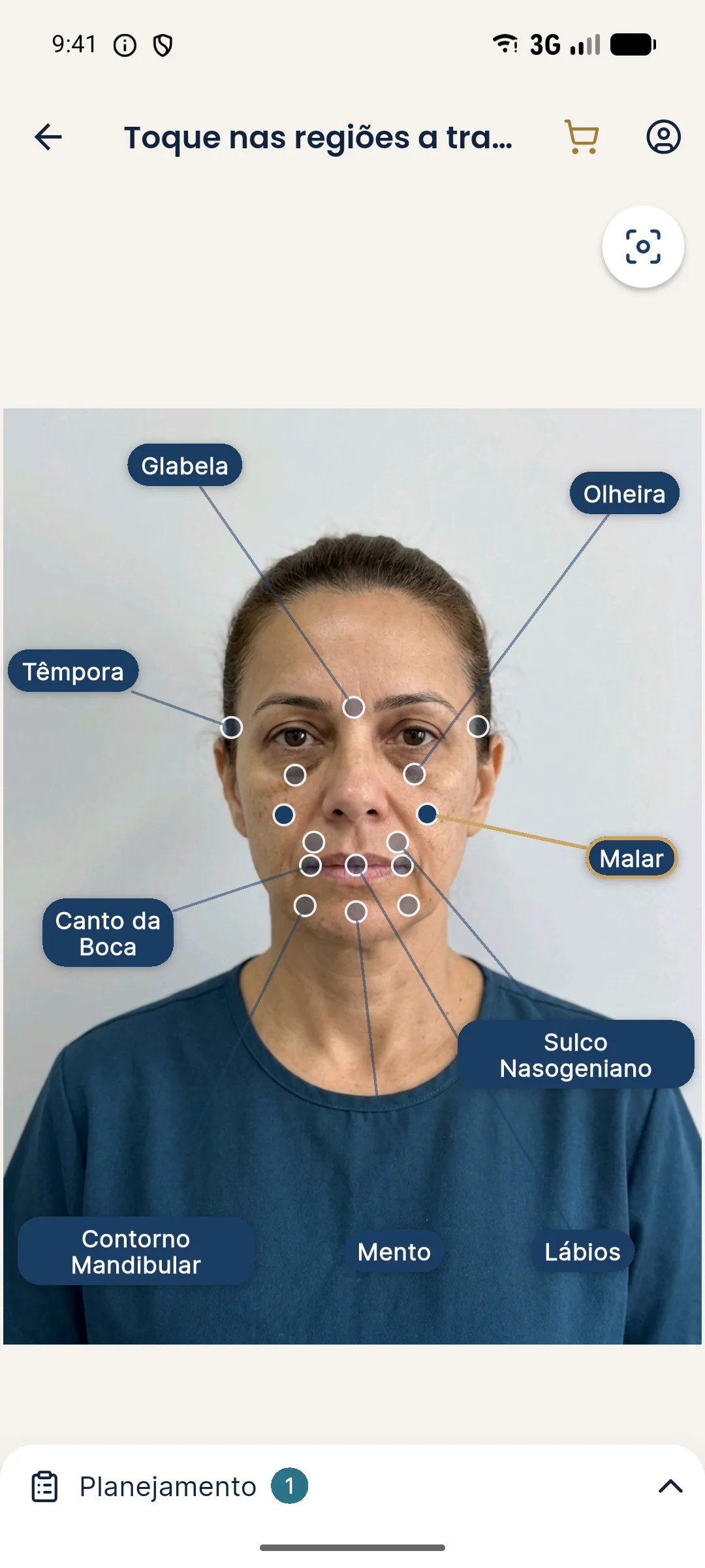
The simulation helps illustrate what changes when the malar area gains support. In Harmy, the projection uses only the front photo and shows the volume result subtly. It does not reposition tissue and does not show the effect of gravity or movement, and it is good for the patient to know this.
When to think about surgery or another path
There are situations where volume only moves the problem around:
- Excess skin or tissue in the cheek, with pronounced descent.
- Descent that already reaches the neck and the jawline, with a marked platysmal band.
- A patient who expects a "lifting" effect from filler.
- Very lax tissue, where the product would tend to slip or become obvious.
In those cases, the honest path is to explain the limit and, if appropriate, refer. For a list of concerns outside the scope, see concerns HOF does not solve, and for the workflow, referring patients.
How to record and follow up
Standardize the photos: same distance, same light, neutral expression, and hair pulled back. Note the predominance you identified (descent, volume, or both), the plan agreed in ranges, and what was said about the limits. At each follow-up, compare at the same angle, because the cheek changes with light and posture. The page standardized clinical photography details the method, and the history per patient keeps sessions organized.
If the patient has recent weight loss, the reading changes: see facial volume loss after weight loss. When the descent reaches the jawline, continue to laxity along the face contour.
Frequently asked questions
What is midface ptosis?
It is the descent of the soft tissues between the eyebrows and the mouth, with the cheek fat migrating downward and forward. The result is usually a deeper fold, a lower cheek, and a more obvious under-eye hollow.
Are ptosis and volume loss the same thing?
No. In volume loss, the tissue shrinks and support decreases. In ptosis, the tissue descends because ligaments and layers lose their hold. Many patients have both, with one predominating.
Does cheek filler lift a sagging cheek?
It can improve the sense of support and the apparent position of the cheek when there is volume loss. With marked descent, the effect tends to be limited, and the indication is up to the licensed professional after assessment.
At what age does midface descent appear?
There is no set age. Signs are generally noticed from about 35 to 45, with wide variation by genetics, weight, sun exposure, and bone structure.
When should surgery be considered?
When the descent is pronounced and there is excess skin or tissue, so that volume would only move the problem around. In those cases it is worth discussing a referral to a surgeon.
Harmy images are illustrative simulations generated by AI from the patient's photo to support the conversation between practitioner and patient. They are not a promise of results. Indication, technique, product and volume are solely the decision of the licensed practitioner. This content is for information only and does not replace a clinical assessment.