
Asymmetric cheeks: layer-by-layer assessment and plan
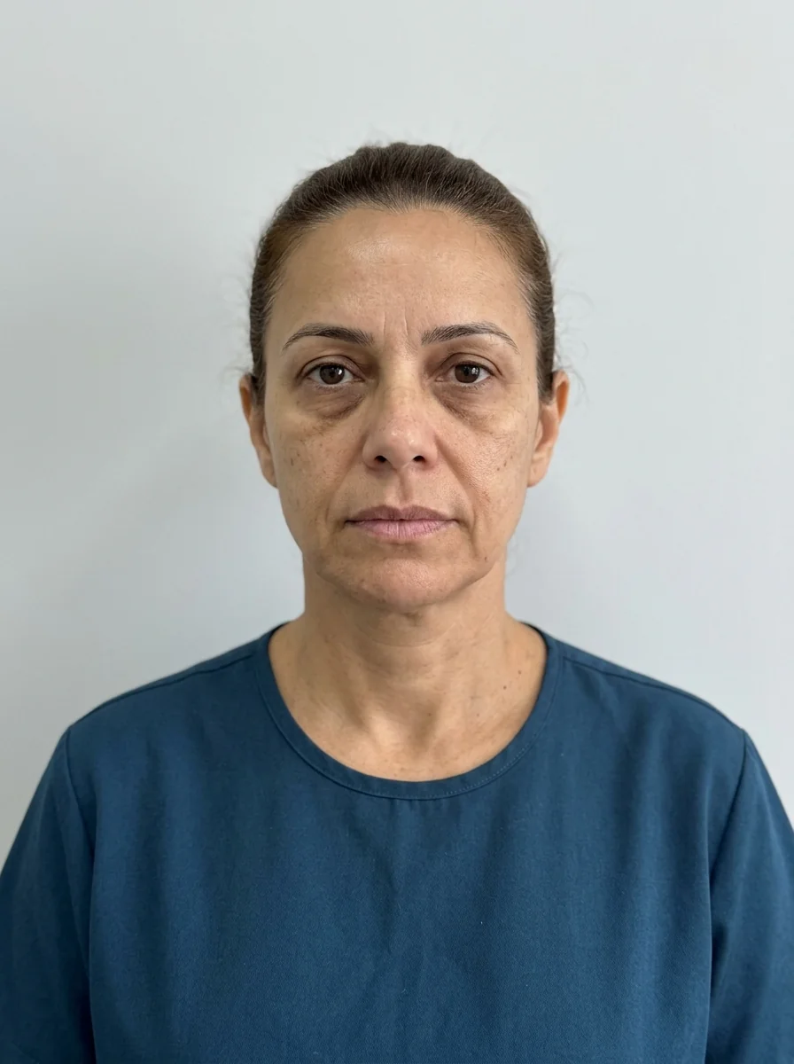
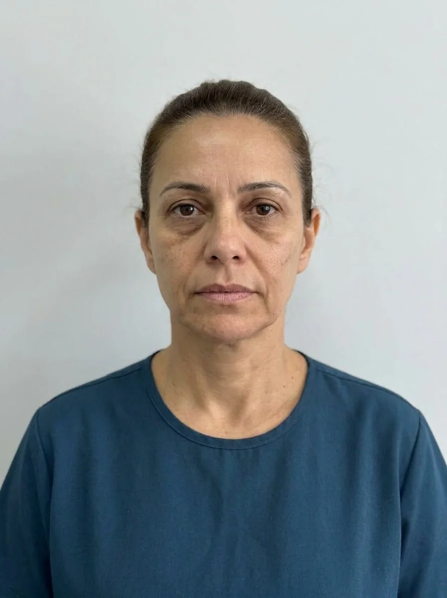 BeforeSimulation
- Highlighted area
Quick facts
- Concern
- One cheek fuller, higher or more projected than the other
- Layers
- Bone, fat, muscle, skin and head posture
- Useful test
- Photos from above and below, palpation and a photo with the head corrected
- Principle
- Correct the difference without chasing perfect symmetry
The cheek is the part of the face where asymmetry is most noticeable in a front-facing photo, because light hits the apple of the cheek and highlights any difference in projection. The patient points and says: "this side is higher" or "this one is fuller." To answer with confidence, you need to know what is different: the bone, the fat, the muscle, the skin or the position of the head.
This page offers a layer-by-layer assessment guide and the logic of the plan. It links to asymmetric face, perfect symmetry is not the goal and plan for an asymmetric face.
Which layers can differ?
| Layer | What can differ | Does facial harmonization change it? |
|---|---|---|
| Bone | Projection and height of the zygomatic bone and the arch | No; only with a soft tissue camouflage plan |
| Fat | Volume of the malar compartments, such as the SOOF and the prezygomatic space | Yes, in part |
| Muscle | Strength and volume of the masseter and the zygomatic muscles | Yes, with toxin in selected cases |
| Skin and ligaments | Tension, laxity, scarring | Partially |
| Posture and habits | Sleeping on one side, tilting the head, chewing on one side | Guided, not injected |
Bone asymmetry is the most common and the least obvious: everyone's face has a more projected side. What turns asymmetry into a problem is the sum of layers that accentuate the difference. See facial asymmetry for the conceptual basis.
How to assess, step by step
The exam needs to be systematic, with standardized photos.
A photo trick: mirroring the right half and the left half of the face (two images: right + mirrored right, left + mirrored left) shows how the face would look if each side were symmetric. Patients are often surprised, and the conversation about what is natural gets easier. Use it with care and with consent, so it does not suggest there is a "correct" face.
What harmonization proposes
The logic is to compensate for the difference without creating a new asymmetry, at the discretion of the qualified practitioner.
- Fat difference: malar filler in a larger amount on the side with less projection, in stages, with reassessment at each follow-up.
- Bone difference: soft tissue camouflage can reduce the perception, but it does not change the bone. Document this limitation.
- Muscle difference: toxin in the masseter and other muscles may be considered in selected cases. See asymmetry from chewing on one side.
- Skin and laxity difference: biostimulation can be added for quality, but it does not resolve a projection difference on its own.
- Posture and habits: give guidance on sleeping on one side and on chewing. See pillow face.
Product planning in an asymmetric face follows the logic of different volumes per side. This runs against what the app does by default, which splits the total ml equally between both sides, so the verbal explanation to the patient is part of the process.
Risks
Risks include swelling, bruising, temporary asymmetry, nodules and irregularities, and vascular complications are less frequent but serious. The area has branches of the transverse facial artery, and the proximity of the eye demands care. Hyaluronidase should be on hand when hyaluronic acid is used. With asymmetry, uneven swelling in the first days can give the impression of getting worse: assess within the settling window. See asymmetry after filler.
How Harmy helps
In Harmy, the Cheekbone area lets you simulate cheekbone support on the frontal photo. In a bilateral area, the ml entered is the total, split between both sides, which means the app does not distribute different volumes per side. For an asymmetry, the most honest use is to show the overall result and explain verbally the difference you intend to compensate for.
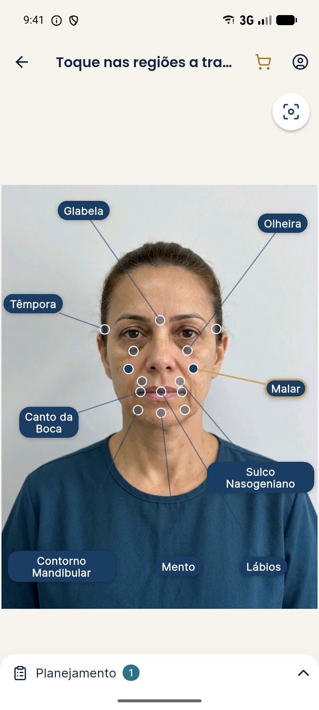
A useful feature is the sample patient, the fictional person who lets you test the flow without a real patient. You can use it to practice explaining the asymmetry before the actual consultation. The projection uses only the frontal photo and changes only what was marked.
How to track progress
Asymmetry is treated in stages, and follow-up is part of the treatment. Agree on a schedule of follow-ups with the patient and always photograph under the same conditions: same distance, same light, same head position and same expression. Without that, comparing sessions is just impression.
A simple table in the patient record, with the date, the volume per side and the patient's assessment, helps reveal the pattern. If the patient always perceives one side as smaller, even after different volumes, it is worth reviewing whether the concern is anatomical or perceptual. It is also useful to note when uneven swelling alters the assessment. Document what was agreed about next steps and maintenance. The goal is for the patient to see the progress and understand that the target is harmony, not sameness between sides.
Wrap-up
Record the standardized photos, the dominant layer and the staged plan. Document the conversation about symmetry as a realistic goal. For cases where the whole face is asymmetric, see simulation for a patient with asymmetry. Volumes and techniques are at the discretion of the qualified practitioner, according to the rules of their professional board.
Frequently asked questions
Can every asymmetric cheek be corrected?
Not entirely. Small differences are normal, and perfect symmetry is not the goal. Soft tissue volume differences can be balanced, but bone asymmetries have a limit with injectables.
How can I tell if the cheek asymmetry is bony?
Palpating the zygomatic arch and the body of the bone, taking a photo from above and comparing with older photos all help. If in doubt, imaging may be considered, at the practitioner's discretion.
Can head posture mimic asymmetry?
Yes. Tilting or rotating the head changes the light and how the volume reads. Standardizing the photo with the head in the Frankfurt plane avoids misreadings.
Does filling only one side solve it?
It can be the strategy, but it takes care not to create one side that is fuller than the other in a different way. The plan is often deliberately asymmetric and reassessed in stages.
Harmy images are illustrative simulations generated by AI from the patient's photo to support the conversation between practitioner and patient. They are not a promise of results. Indication, technique, product and volume are solely the decision of the licensed practitioner. This content is for information only and does not replace a clinical assessment.