
Ptosis or Volume Loss: Which to Treat First in the Plan

- Muscle
- Highlighted area
Quick facts
- Central question
- Is the face sagging (ptosis) or hollowing (volume loss)? Often both, in different proportions
- Simple test
- Lift the tissue by hand and observe whether volume reappears or whether there is excess skin and sagging
- Strategies
- Replace volume, support the tissue, stimulate collagen or combine, at the professional's discretion
- Caution
- Replacing volume in someone with mainly ptosis can weigh the face down; supporting without replacing can leave the face gaunt
When a patient says "my face is sagging", they are talking about two very different things under the same concern. One is the descent of tissue, ptosis. The other is hollowing, volume loss. In clinical practice, the two coexist, in different proportions. Identifying which one predominates is one of the most important steps in the plan, because it determines whether the strategy will be to replace, support, stimulate or combine.
This page offers an assessment reasoning, at review level, to decide what comes first. It does not describe injection technique, and the final approach belongs to the qualified professional, after assessment, according to the rules of their professional board.
What sets ptosis apart from volume loss?
Ptosis is the descent of soft tissue. Skin and fat are still there, but they have slid, because ligaments and support structures have loosened. Traction from their own weight and gravity, over the years, favors displacement. The structures involved are in retaining ligaments, SMAS and fat pads.
Volume loss is an actual reduction of tissue: fat (superficial and deep), bone or muscle. Fat compartments shrink and shift, and bone also resorbs over time, as in facial bone resorption and planning. The result is a more hollowed face with sharper contours.
Which clinical tests help decide?
None is definitive. Together, they guide the hypothesis:
- Lift test: with your fingers, gently lift the tissue of the cheekbone and cheek area. If the contour improves noticeably, there is a ptosis component. If the tissue returns without changing the hollow look, the volume component is greater.
- Jawline: loss of definition with buildup below the line (jowl) suggests ptosis, covered in jowl. A well-defined line with depressions suggests volume loss.
- Malar plane: a flat cheekbone with tissue shifted downward suggests ptosis. A flat cheekbone with absent tissue suggests volume loss.
- Folds: deepening of the nasolabial fold and the pre-jowl sulcus has its origin in both. The reasoning is in treating the fold or the cause of the fold.
- Skin: laxity and excess skin weigh toward ptosis. Thin, hollowed skin weighs toward volume loss. Thickness appears in skin thickness in planning.
| Sign | Points more to ptosis | Points more to volume loss |
|---|---|---|
| Lift test | Clear improvement | Slight improvement |
| Jawline | Jowl, loss of definition | Defined line, depressions |
| Cheekbone | Flat with descended tissue | Flat with absent tissue |
| Skin | Excess and laxity | Thin, hollowed |
| Standing and lying photos | Changes a lot | Changes little |
How does the predominance change the strategy?
The predominance guides the type of intervention, and the set of support and replacement strategies appears in volumization and support.
- Predominant volume loss: replacing volume in structural areas is usually the base, taking care not to exceed what bone and skin can support.
- Predominant mild to moderate ptosis: support and stimulation strategies, such as thread lift and collagen biostimulators, are described alternatives. The role of volume is limited.
- Predominant marked ptosis: the role of injectable procedures is smaller, and the conversation with the patient should include other options, including surgical ones, and when to refer.
- Coexistence: the combination is common and requires a sequence, as in threads and filler: order in the plan and in biostimulator and filler: combining areas.
What changes in each area?
In the cheekbone, volume loss usually calls for structural replacement, as in malar filler. The descent of malar tissue, on the other hand, calls for reading the whole, since a cheekbone projected on its own does not fix what has descended. See ideal malar projection.
In the jaw and jowl, the definition of the line is affected by both. When the jowl dominates, simply adding volume along the jawline may help hide the transition, but the sign is one of ptosis. Jawline filler has a role when there is a contour deficit, while descent has a different logic. The texts jaw and masseter: combining them in the plan and chin and double chin: combining them in the plan go into detail.
In the nasolabial fold, both hollowing and descent of the cheekbone contribute. The area carries high vascular risk, which reinforces the importance of treating the cause first, with hyaluronidase on hand when the product is hyaluronic acid.
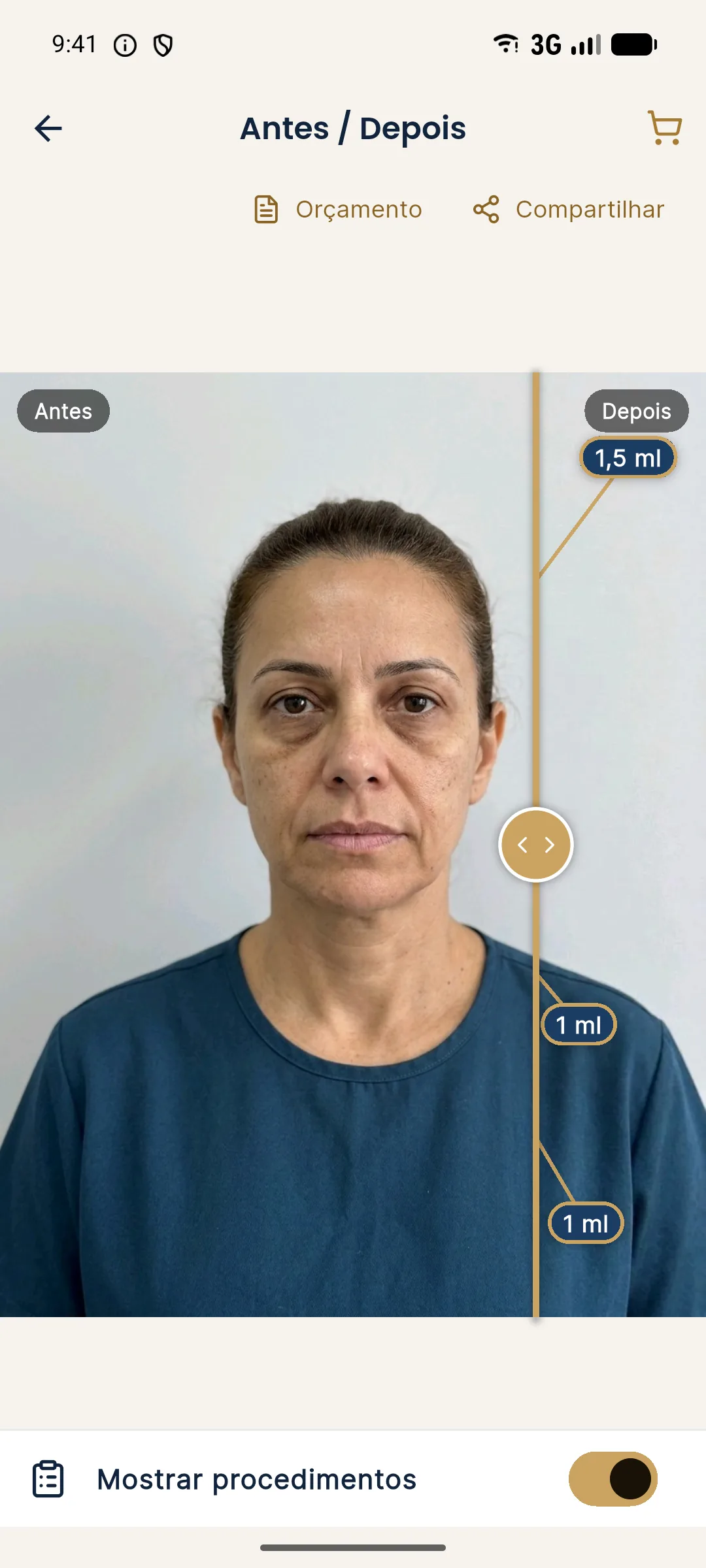
How to record the assessment?
Photograph standing, seated and, if possible, lying down, always with standardized light and distance. Record the lift test result by area and your hypothesis of predominance. This allows comparison at follow-up. The photo workflow is in photographic record by plan stage and in frontal facial analysis: checklist.
How to explain it to the patient?
Avoid jargon. Use the image of a curtain: it may be hung loosely (ptosis) or made of too little fabric (volume loss). The treatment changes depending on the case. Reinforce that the result is individual, that there are limits and that not every sag is fixed with injectables.
The same clarity applies to the patient who brings an expectation of a surgery-like result: see explaining what facial harmonization does not solve and facial rejuvenation plan.
Frequently asked questions
What is the difference between ptosis and volume loss?
Ptosis is the descent of soft tissue from loosened support. Volume loss is a reduction of fat, bone or muscle. The two can coexist and be confused in the mirror.
How do I know which one predominates?
Clinical assessment looks for clues: the tissue returns to place when lifted, the jawline has lost its line, the malar area is flat or only displaced. Photos in several positions help.
Does filler treat ptosis?
It can improve the look of support in mild to moderate ptosis, at the professional's discretion. In marked ptosis, other strategies, including surgical ones, may be more suitable.
What happens if I treat the wrong problem?
Replacing volume in predominant ptosis can weigh down the lower third and accentuate the jowl. Supporting without replacing, in volume loss, can leave the face gaunt. Identifying the predominance avoids both mistakes.
Harmy images are illustrative simulations generated by AI from the patient's photo to support the conversation between practitioner and patient. They are not a promise of results. Indication, technique, product and volume are solely the decision of the licensed practitioner. This content is for information only and does not replace a clinical assessment.