
Facial Assessment by Thirds in Orofacial Harmonization
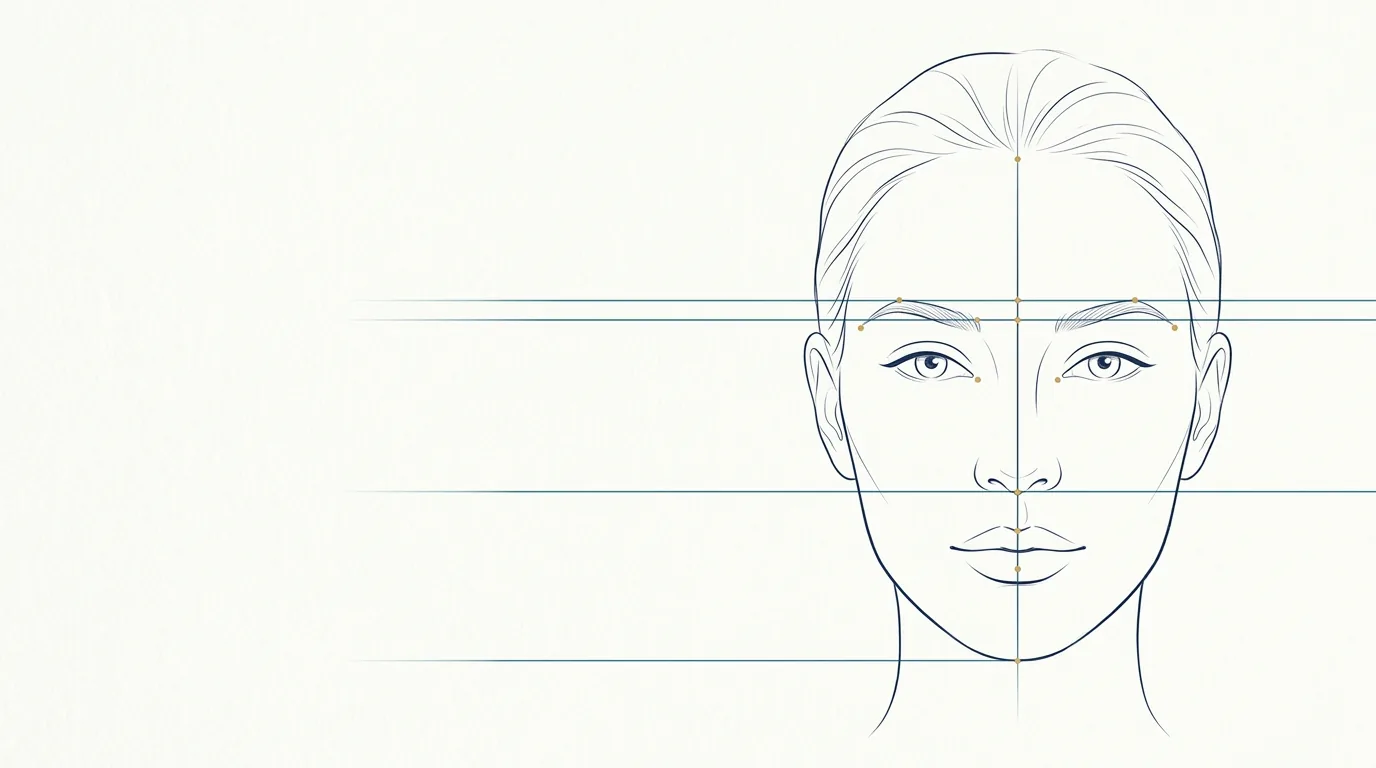
Before thinking about product or volume, orofacial harmonization (HOF) starts with a question: where is the imbalance? Assessment by thirds is the most widespread way to answer it in an organized fashion. It divides the face into comparable bands, helps you see the face as a whole and gives a common language to explain the plan to the patient.
This article presents the method at a review level: horizontal thirds, vertical fifths, profile analysis, reading asymmetries and how all of that becomes treatment priorities.
What are the thirds of the face?
In the frontal view, with the head in the Frankfurt plane, the face is divided by horizontal lines into three bands:
| Third | Usual limits | Main structures |
|---|---|---|
| Upper third | From the hairline (trichion) to the glabella | Forehead, glabella, eyebrows, temples |
| Middle third | From the glabella to the base of the nose (subnasale) | Eyes, periorbital region, cheekbone, zygomatic area, nose, start of the nasolabial fold |
| Lower third | From the base of the nose to the lowest point of the chin | Lips, mouth corners, chin, jaw |
The classic reference considers the three thirds to be of roughly equal height. In practice, that equivalence is a point of comparison, not a goal: the hairline varies a great deal, and individual and ethnic differences are part of faces considered harmonious.
How to subdivide the lower third?
The lower third is usually read in two parts, separated by the line where the lips meet:
- from the subnasale to the lip contact, the upper lip portion, including the philtrum;
- from the lip contact to the chin, the lower lip and chin portion.
The classic reference puts the upper lip at about one third of that height and the lower lip and chin together at about two thirds. A long upper lip, a short chin or an elongated chin quickly show up in this reading.
What about the vertical fifths?
The horizontal assessment is completed by vertical lines that divide the width of the face into five parts. As a classic reference, each fifth is close to the width of one eye:
- from the lateral edge of the face to the outer corner of the right eye;
- the width of the right eye;
- the space between the eyes;
- the width of the left eye;
- from the outer corner of the left eye to the lateral edge of the face.
The fifths help to perceive a narrow or wide face in the middle third, the distance between the eyes and the proportion between nose width and the intercanthal space. They also guide the reading of the temples and zygomatic area, which define the width of the upper and middle thirds.
How to assess the profile?
The frontal view shows height and symmetry, but it does not show projection. For that, assessing the facial profile is indispensable. Points that are usually observed:
- Profile type: straight, convex or concave, based on the relationship between forehead, base of the nose and chin.
- Chin projection: a retruded or projected chin relative to the lips and nose.
- Lip projection: position of the lips relative to a reference line joining the tip of the nose to the chin (Ricketts' esthetic line is the most cited).
- Nasolabial angle: the opening between the columella and the upper lip.
- Jawline: definition of the jawline and the mandibular angle, and the transition between jaw and neck.
A retruded chin, for example, can make the nose look bigger and the lips more projected. Treating the chin changes how all three structures read, which is why the profile often changes the priority of the plan.
How to read asymmetries?
No face is perfectly symmetric. Mild facial asymmetry is the rule, and much of it is skeletal or dental, beyond the reach of HOF. The assessment looks for:
- volume differences between the sides (cheekbone, temples, lips);
- height differences (eyebrows, mouth corners, jaw angles);
- midline deviations (chin, philtrum, nose);
- movement asymmetries, observed in expression (smile, eyebrow lift).
Recording these asymmetries before treating is important for two reasons: adjusting the plan side by side, at clinical discretion, and showing the patient what was already there. Many complaints of "it came out crooked" after a procedure come from pre-existing asymmetries nobody pointed out.
What to observe in each third?
Upper third
- Horizontal forehead lines and glabella lines at rest and in contraction.
- Position and shape of the eyebrows.
- Volume of the temples: lateral hollowing narrows the upper third and accentuates a tired look.
Common procedures: toxin in the glabella, toxin in the forehead lines, temple filler.
Middle third
- Support and volume of the cheekbone and the zygomatic area, seen from the front and in the oblique view.
- Tear trough and under-eye area, including the transition between eyelid and cheek.
- Depth of the nasolabial fold (smile lines), which often reflects loss of support above it.
- Dorsum and tip of the nose, in frontal and profile views.
Common procedures: cheekbone filler, zygomatic filler, tear trough filler, fold filler.
Lower third
- Volume, contour and symmetry of the lips, proportion between the upper and lower lip.
- Position of the mouth corners.
- Projection and height of the chin.
- Definition of the jawline.
Common procedures: lip filler, chin filler, jawline filler, mouth corner filler.
How to turn the assessment into a plan?
The assessment is only worth something if it becomes priorities. A common logic, at the professional's discretion:
- Structure before detail. Support areas (temples, cheekbone, chin, jaw) usually come before folds and lines, because they change how neighboring areas read.
- The most disproportionate third first. If the lower third is short and retruded, working only on the lips tends to accentuate the imbalance.
- Stages, not excess. Volumization over several sessions allows reassessment and lowers the risk of overcorrection.
- Aligned expectations. The patient needs to understand what is structural, what is treatable and what does not change with HOF.
Hypothetical example
Hypothetical example: a patient complains about "smile lines." The assessment shows a cheekbone with little projection, a deep fold and a slightly retruded chin in profile. Instead of treating only the fold, the plan discussed includes cheekbone support and chin projection in stages, with the fold reassessed afterward. Volumes and technique are at the professional's discretion, after examination.
Where does Harmy fit in the assessment?
Harmy does not do the assessment for you. It helps show the patient the plan that came out of it. After a frontal photo, the app detects the face on the device itself and shows the points of the areas, distributed across the three thirds: Glabella and Temple in the upper; Tear trough, Cheekbone and Nasolabial fold in the middle; Mouth corner, Lips, Chin and Jawline in the lower.
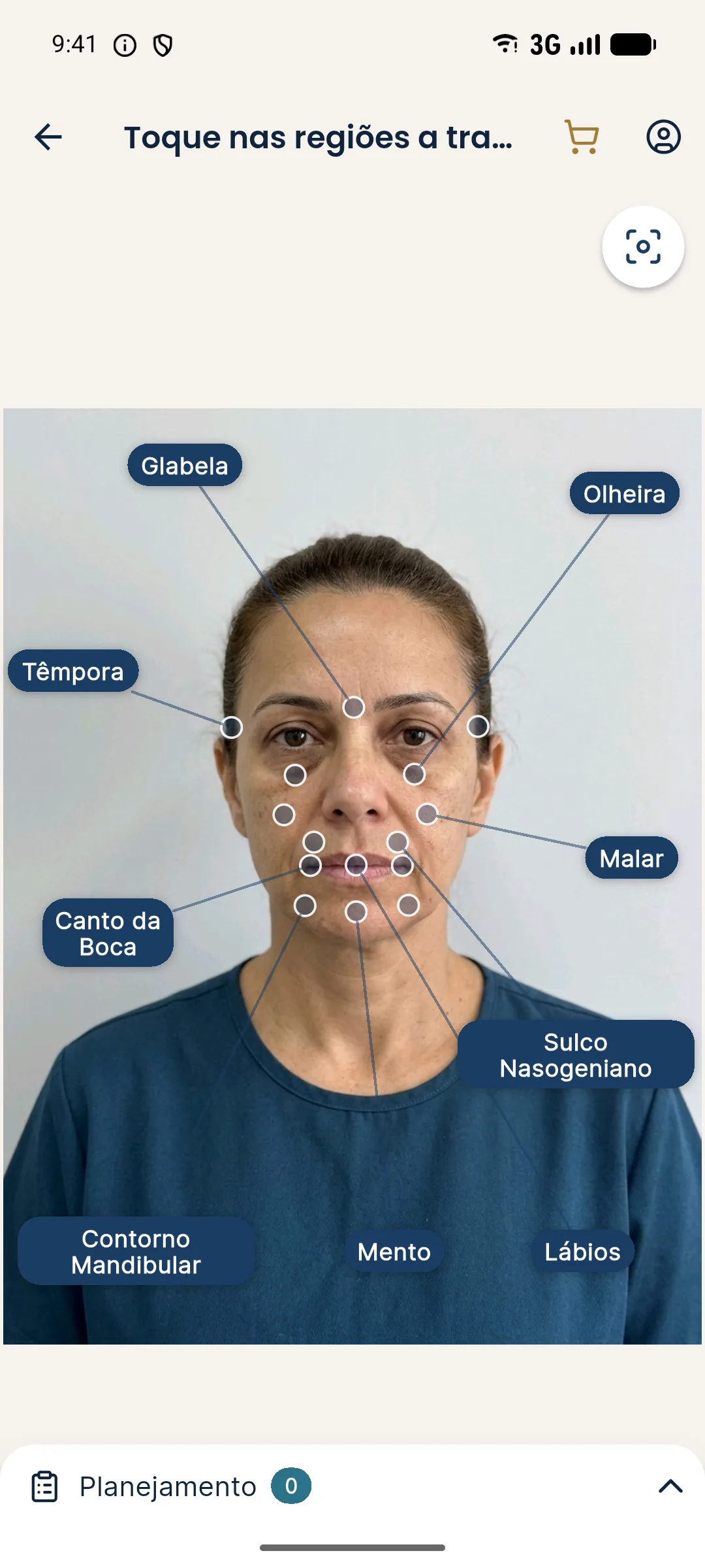
You mark the areas in the plan, set the volume of each in ml (from 0.5 to 5 ml) and generate the projection. Because the image changes only the marked areas, it works well to show the effect of treating the structure, for example comparing a plan with only the fold against one with cheekbone and chin. Remember that the projection is an illustrative simulation, made on the frontal view: it does not show the profile and is not a promise of results.
For the input photo, follow the guide to standardized clinical photography. For the full map of areas, see the overview of treatment areas.
Checklist for assessment by thirds
| Step | Done |
|---|---|
| Standardized photos: frontal, oblique and profile views | |
| Heights of the three thirds compared | |
| Lower third subdivided (upper lip vs. lower lip and chin) | |
| Vertical fifths and width of the middle third | |
| Profile: type, lip and chin projection, jawline | |
| Asymmetries noted and shown to the patient | |
| Expressions assessed (forehead, glabella, smile) | |
| Priorities and stages defined | |
| Risks discussed and consent recorded |
Assessing by thirds is not about fitting the patient to a ruler. It is about having a script for looking at the whole face, explaining what you see and proposing a plan consistent with the concern, the anatomy and safety. On the safety side, see the overview of complications.
Frequently asked questions
What is facial assessment by thirds?
It is the division of the face into three horizontal bands (from the hairline to the glabella, from the glabella to the base of the nose and from the base of the nose to the chin) to compare their heights and characteristics. It serves as a starting point, not as a rigid rule.
Do the three thirds of the face have to be equal?
No. Roughly equal thirds are a classic reference, but individual and ethnic variation is normal. The assessment looks for imbalances that bother the patient or that treatment can soften, not mathematical equality.
Which procedures work on each third?
In the upper third, botulinum toxin and temple filler predominate; in the middle, cheekbone, zygomatic and tear trough filler; in the lower, lips, chin, mouth corners and jawline. The choice is always at the professional's discretion.
Can the thirds be assessed from the front photo alone?
The frontal photo shows heights and symmetry, but not projection. The chin, lips, nose and jaw also depend on the profile. That is why clinical assessment uses frontal, oblique and profile views.
Harmy images are illustrative simulations generated by AI from the patient's photo to support the conversation between practitioner and patient. They are not a promise of results. Indication, technique, product and volume are solely the decision of the licensed practitioner. This content is for information only and does not replace a clinical assessment.