
Zygomatic region (zygomatic arch): anatomy and filler
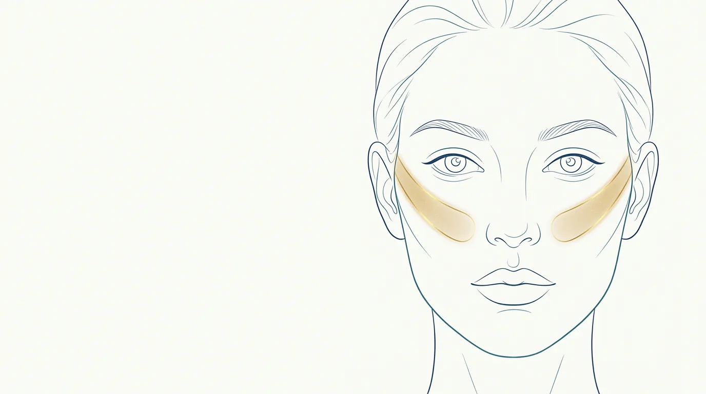
The zygomatic region corresponds to the zygomatic arch, the bony bridge that runs from the cheekbone toward the ear. It defines the width and the lateral contour of the midface. While the malar area is the most anterior part of the cheekbone, the zygomatic area is the lateral "line" that gives the face its shape in frontal and three-quarter views.
What are the anatomical boundaries of the zygomatic region?
The zygomatic arch is formed by the temporal process of the zygomatic bone and the zygomatic process of the temporal bone. The practical boundaries of the region are:
- Anterior: the malar eminence, where the region continues into the malar area.
- Posterior: the preauricular region.
- Superior: the temporal fossa, where the temples begin.
- Inferior: the cheek and the masseter region.
Over the arch lie the skin, the subcutaneous tissue, the musculoaponeurotic layer and the fasciae, as well as the origins of the zygomatic muscles and the upper portion of the masseter, which inserts on the lower border of the arch.
Vessels and nerves to watch
- Zygomatic and temporal (frontal) branches of the facial nerve, which cross the arch toward the orbit and the forehead.
- Transverse facial artery, which runs below the arch, parallel to it.
- Superficial temporal artery and vein, in the posterior portion, near the ear.
- Zygomaticoorbital artery and zygomaticofacial branches.
- Parotid duct, below the arch, heading toward the cheek.
Injury to the temporal branch of the facial nerve can affect brow elevation. The posterior portion of the arch, near the temporal vessels, calls for extra attention.
What do patients usually ask for?
Concerns rarely mention the arch:
- "I want a more defined face, like a model."
- "My face is very round."
- "I'd like higher cheekbones."
- "I lost my contour after I lost weight."
Assess the patient from the front, in three-quarter view and with side light. The projection of the arch, the bizygomatic width and the relationship with the jaw guide the indication. In faces that are already wide in the midface, enhancing the zygomatic area can unbalance the proportion.
What procedures can be done in the zygomatic area?
The main procedure is zygomatic filler, with high G' hyaluronic acid, suited to contouring and definition. Some professionals use calcium hydroxylapatite for the same purpose.
The zygomatic area is almost always planned together with:
- Cheekbone filler, for the anterior portion.
- Temple filler, to soften the transition between the upper third and the midface.
Technique, plane and the choice of cannula or needle are the decision of the qualified professional, according to the rules of their professional council in Brazil.
What are the usual volumes in the zygomatic area?
Usual ranges in ml of hyaluronic acid, at the discretion of the qualified professional, after assessment:
| Goal | Usual range per side | Note |
|---|---|---|
| Subtle contour enhancement | 0.5 to 1 ml | Common in young patients |
| More defined contour | 1 to 1.5 ml | Assess face width |
| Volume loss with age | Combine cheekbone and temples | Integrated midface treatment |
| Durability | Generally 12 to 18 months | Varies with product and patient |
Small changes to the arch greatly alter how the face reads. That is why a conservative approach, with follow-up, tends to be the safest.
Precautions, contraindications and complications
Avoid or postpone in the presence of active infection, pregnancy or breastfeeding, uncontrolled autoimmune disease, allergy to the components or unrealistic expectations. Note any medications that increase bleeding risk.
Reported complications:
- Swelling and bruising.
- Excessive widening of the face or an artificial look.
- Facial asymmetry between the sides.
- Palpable irregularity over the bone.
- Vascular occlusion, with risk of necrosis, especially near the temporal vessels.
In the zygomatic region with hyaluronic acid, keep hyaluronidase ready for the appointment. The overview of risks is in safety and complications.
How to assess the zygomatic arch
Since small changes to the arch alter how the face reads so much, the assessment must consider overall proportion:
- Bizygomatic width relative to the jaw: faces in which the midface is already much wider than the lower third call for caution.
- Side light: highlights the shape of the arch and shows whether there is a hollow below it.
- Three-quarter view: this is where the lateral contour shows most clearly.
- Transition with the temples: a temporal concavity above the arch can make the zygomatic area look more prominent than it is.
- Patient's goal: someone who asks for a "defined" face does not always want a wider face.
Facial assessment by thirds helps place the midface within the whole face. Photographing the patient always with the same light and the same framing makes it easier to compare the contour at follow-up, since small changes to the arch are hard to see with the naked eye. See the guidelines on standardized clinical photography.
How does the zygomatic area appear in Harmy?
The zygomatic area has no separate point in Harmy's marking. In the app's catalog, zygomatic filler is represented by the Cheekbone point, the same one used for the cheekbone area.
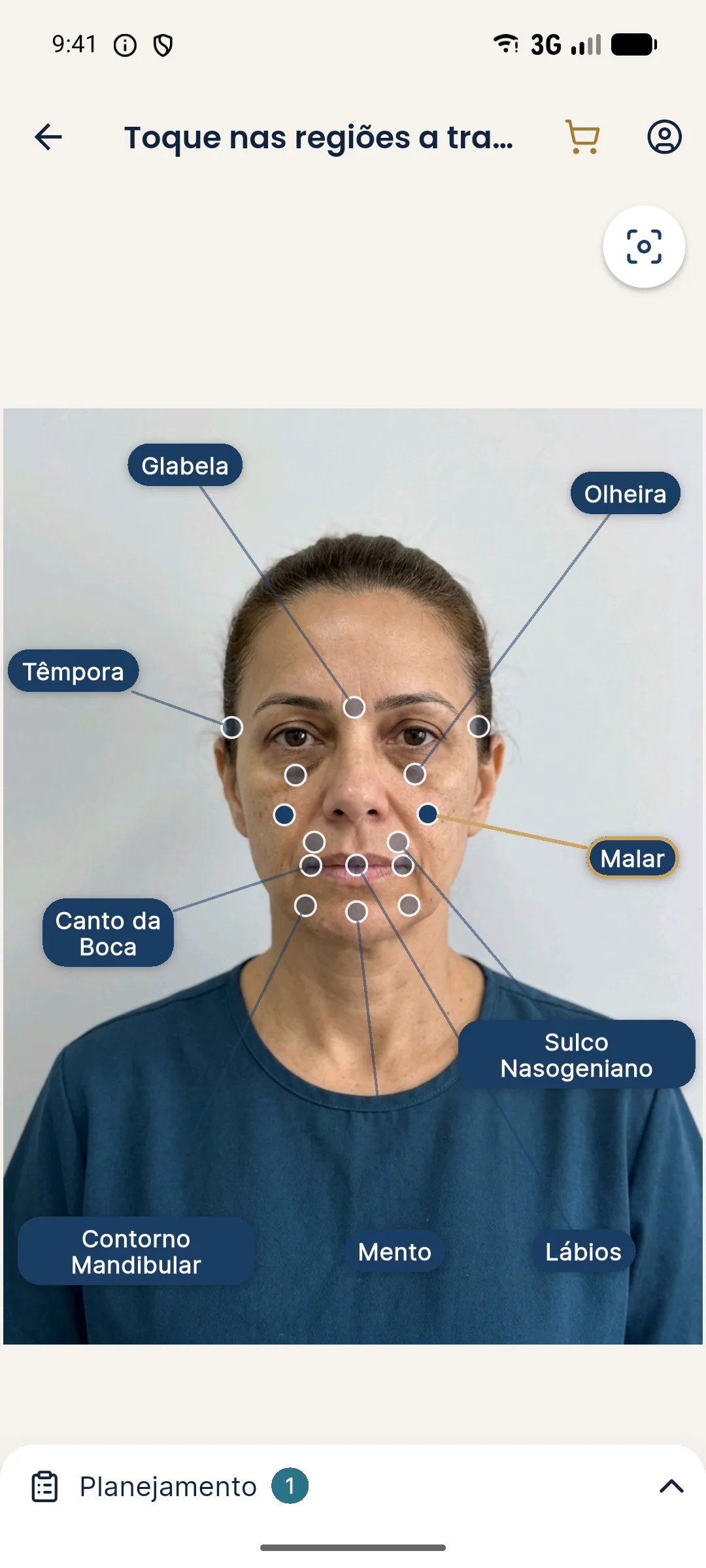
In practice, when you mark the Cheekbone and set the volume in the Plan (0.5 ml steps, from 0.5 to 5 ml), the projection shows the gain in support and contour in the midface. Because it is a bilateral area, the value entered is the total, split between the two sides. The AI changes only what was marked, with no skin retouching, makeup or change of expression.
See the step by step in marking areas and volumes.
How to talk to the patient about the zygomatic area
Someone who asks for a "defined face" often has a social media reference in mind. Showing an illustrative simulation on the patient's own photo brings the conversation back to their face, with real proportions, and helps align how much contour makes sense.
Use the projection as support, make clear it is not a promise, and agree on the plan in stages when appropriate. To lead that conversation, see how to present before and after in the consultation.
Frequently asked questions
What is zygomatic filler?
It is the application of filler along the zygomatic arch to enhance the lateral contour of the midface. It is usually planned together with the cheekbone and the temples.
How much filler is used in the zygomatic area?
Generally 0.5 to 1.5 ml per side, at the discretion of the qualified professional after assessment. The amount depends on face shape and the contouring goal.
Does zygomatic filler make the face wider?
It can, if the volume or position is not suited to the shape of the face. That is why the assessment of proportions comes before the indication.
Does Harmy have a point for the zygomatic area?
Not a separate point. In the app, zygomatic filler is represented by the Cheekbone point, which covers the cheekbone area.
Harmy images are illustrative simulations generated by AI from the patient's photo to support the conversation between practitioner and patient. They are not a promise of results. Indication, technique, product and volume are solely the decision of the licensed practitioner. This content is for information only and does not replace a clinical assessment.