
Profile Facial Analysis: What to Assess and Measure
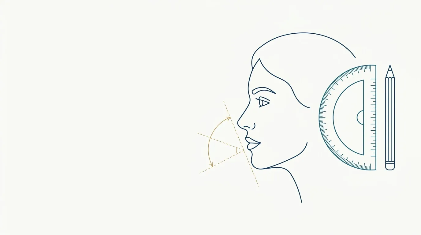
- Nerve
- Highlighted area
Quick facts
- Goal
- Read the relationship between forehead, nose, lips, chin and neck in the lateral view
- Prerequisite
- True profile photo with the head in the Frankfurt plane
- Usual references
- Steiner S line, Ricketts line, H line and facial angles
- Limit
- Measurements are a guide, not a goal; the patient and the individual bone rule
The frontal view shows width and symmetry, but it is the profile that reveals projection. A recessed chin, a retracted lower lip, a jawline that does not define itself: all of this is clearer from the side. Many complaints that the patient attributes to a "small chin" or a "big nose" are, in fact, a relationship between structures that only the profile shows.
This page is a checklist for reading the lateral view for the facial harmonization plan. It covers photo positioning, reference lines and angles, the step-by-step reading and the mistakes that most distort the assessment. For the overview of the assessment, see facial analysis in facial harmonization: step by step and facial assessment by thirds.
How to photograph the profile so the analysis is valid?
Without a standardized photo, any measurement is noise. The head must be in the Frankfurt plane: an imaginary line running from the upper edge of the external ear canal to the lowest point of the orbit, parallel to the floor. The gaze goes to the horizon, the lips are relaxed and the teeth in habitual occlusion.
Details on hair, earrings and glasses are in hair, earrings and glasses in the assessment photo, and the choice of lens and distance in distance and lens in the assessment photo. The complete photo routine is in standardized clinical photography.
Which lines and angles organize the reading?
Lines and angles help name what the eye already perceives. They are not goals: they vary with sex, ancestry and body type, and the patient's face can be well balanced even outside a published range.
| Reference | What it relates | What it is for in the plan |
|---|---|---|
| Steiner S line | Lips relative to a line between the contour of the nose and the chin | Assess lip and chin projection |
| Ricketts line | Tip of the nose to the chin (esthetic plane) | See lip position relative to the nose and chin axis |
| Holdaway H line | Upper lip to the chin | Assess harmony of the lip with the chin |
| Nasofrontal angle | Forehead and nasal dorsum, at the nasion | Read the root of the nose |
| Nasolabial angle | Columella and upper lip | Read tip rotation and upper lip projection |
| Mentolabial angle | Lower lip and chin | Read fold depth and chin projection |
| Cervicomental angle | Submentum and neck | Read the double chin and neck definition |
| Facial convexity angle | Glabella, subnasale and pogonion | Classify a straight, convex or concave profile |
Reference values vary by author, population and method. Instead of memorizing ranges, use them as a question: "is this lip further forward or back than the whole suggests?". The discussion on ideals is in does a facial aesthetic ideal exist? and in facial proportions vary between populations.
In what order should the profile be read?
A fixed sequence avoids skipping steps and fixating on a single area.
- Forehead and glabella: forehead slope, brow ridge and root of the nose.
- Nose: dorsum, tip, columella and nasolabial angle.
- Lips: projection of the upper and lower lip, relative to a reference line.
- Mentolabial fold and chin: fold depth and position of the pogonion. An evident recession of the chin relative to the lips appears in retrogenia.
- Jawline and submentum: definition of the angle and transition to the neck.
- Overall: whether the profile looks convex, straight or concave, and what causes it.
Where does the profile change the plan?
The profile usually changes the priority more than the list of areas. Two well-known examples:
- Recessed chin: the patient complains about a big nose or a thin lip, but the problem is the lack of chin projection. Correcting the base changes how the rest is perceived. See plan to project the chin and ideal chin projection by sex.
- Lips and nose together: the tip of the nose, its rotation and the upper lip form a unit. See aesthetic profile: lips, nose and chin and nose and upper lip: combining in the plan.
For those seeking overall balance, plan to balance the profile gathers the decision logic, and maxillary projection and midface in the profile covers support of the midface. When the problem is bone, remember when bone is the problem, not volume.
And Harmy, where does it come in?
The limitation is useful to explain to the patient: the projection shows from the front what the plan intends to change, but does not show what happens from the side. If the plan depends on chin or lip projection, say that the simulation does not capture the profile change. More in limits of 2D simulation.
What ethical care applies to profile analysis?
The profile is a photo of the face and therefore sensitive personal data: ask for specific consent to photograph, store and use the record, and avoid publishing it without written authorization. See consent and LGPD in photos (LGPD is Brazil's data protection law). Lines and angles should not be used to create insecurity in the patient ("your chin is outside the range"), but to organize the conversation.
Record in the patient record the head position, the distance and the references used, so the next photo can be compared with the same ruler. Every indication of product, volume and technique belongs to the licensed professional, according to the rules of their professional council.
Frequently asked questions
What should be assessed in profile facial analysis?
Forehead, nose, lip projection, chin, mentolabial angle, jawline and the transition to the neck. The reading combines angles and reference lines with clinical judgment.
How should the head be positioned in the profile photo?
With the Frankfurt plane parallel to the floor, eyes on the horizon, lips relaxed and teeth in habitual occlusion. A tilted head changes the reading of the chin and the nose.
Are profile measurements rules I have to follow?
No. They are reference ranges used as a guide, and they vary with sex, ancestry and individual characteristics. The plan starts from the patient's face, not from a number.
Does Harmy simulate the patient's profile?
No. Harmy's projection uses only the frontal photo. The profile assessment is still done by you, in the consultation and in clinical photography.
Harmy images are illustrative simulations generated by AI from the patient's photo to support the conversation between practitioner and patient. They are not a promise of results. Indication, technique, product and volume are solely the decision of the licensed practitioner. This content is for information only and does not replace a clinical assessment.