
Ecchymosis: bruising after injectables vs. hematoma
Quick facts
- Definition
- A flat, purplish patch caused by blood leaking from small vessels and spreading through the tissue.
- Difference from a hematoma
- In ecchymosis the blood stays diffuse, without forming a collection; in a hematoma it pools and forms a palpable lump.
- Color progression
- Purplish or bluish at first, then greenish and yellowish, until it fades.
- Usual duration
- Generally a few days to two weeks, depending on the area and the patient.
- Warning sign
- A lacy patch that spreads, with disproportionate pain or pallor, is not ordinary ecchymosis.
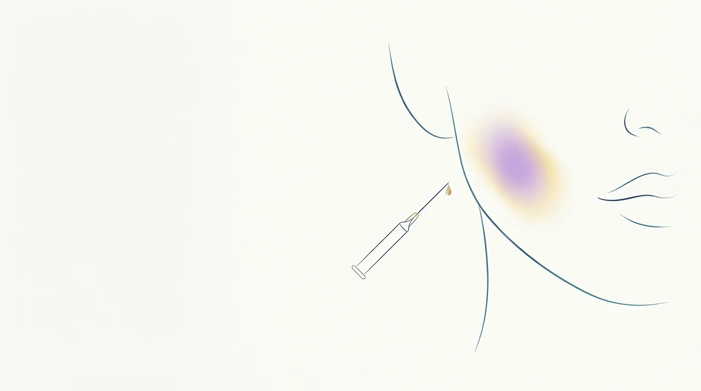
Ecchymosis is the flat, purplish or bluish patch that appears when blood leaves small damaged vessels and infiltrates the tissue diffusely. In orofacial harmonization, it is the "bruise" that may appear at the puncture point after filler, botulinum toxin or other injectables. It is one of the most common complications and, in the vast majority of cases, it is short-lived and only cosmetic.
What is the difference between ecchymosis and a hematoma?
Both come from the same event, damage to a vessel by the needle or cannula, but the blood behaves differently:
| Feature | Ecchymosis | Hematoma |
|---|---|---|
| How the blood sits | Spread between the tissue fibers | Pooled in a collection |
| To the touch | Flat, no volume | Raised area or palpable lump, sometimes tender |
| Size | From a small patch to a larger area | Variable, with volume |
| Course | Fades as the pigment is reabsorbed | May take longer, because the collection has to be reabsorbed |
In practice the terms get mixed up, and many people call any bruise a hematoma. The page on hematoma details the collection. In dermatology, small pinpoint spots have other names, such as petechiae.
Why does ecchymosis change color?
The color follows the breakdown of hemoglobin in the tissue. At first the patch is reddish or purplish; then it turns bluish and dark; as pigments derived from hemoglobin form, it becomes greenish and, finally, yellowish or brownish, until it disappears. In thin-skinned areas, such as around the eyes, the patch can look darker and last longer. Gravity pulls blood downward, so ecchymosis may appear a few centimeters below the injection point.
What increases the risk?
- Highly vascular areas, such as the lips and the periorbital area.
- Number of punctures: each entry of the needle is a chance to damage a vessel. The cannula usually reduces that number, without eliminating the risk.
- Medications and supplements that interfere with clotting, along with the patient's medical conditions. The medical history should ask about them, and no prescribed medication should be stopped without guidance from the patient's physician. The topic is covered in anticoagulants and injectable procedures.
- Thin skin, capillary fragility and age.
How do you tell it apart from a warning sign?
Ordinary ecchymosis appears at the puncture point or just below it, has defined edges and does not come with disproportionate pain. It should also not be confused with the Tyndall effect, which is bluish, stable and does not change color over the days.
How should the patient be advised?
Warning them beforehand avoids a scare and helps the patient not to schedule the procedure close to an important event. Local compression right after the puncture helps limit bleeding. Photographing the patch at the follow-up also documents its course and helps tell it apart from other complications. The conversation about bruising on the lips is in bruising after lip filler.
Also called: bruise, purple patch, ecchymosis.
Frequently asked questions
What is the difference between ecchymosis and a hematoma?
In ecchymosis, the blood that left the vessel infiltrates the tissue and forms a flat patch. In a hematoma, the blood pools in one spot and forms a raised area or a lump you can feel on touch. Both change color over the days.
Is bruising normal after filler?
A well-defined purple patch at the injection point is usually ecchymosis, common and short-lived. A net-like patch that spreads, with severe pain, pallor or cold skin, may be a sign of vascular occlusion and requires immediate assessment.