
Lower Eyelid: Anatomy and the Eyelid-Cheek Transition
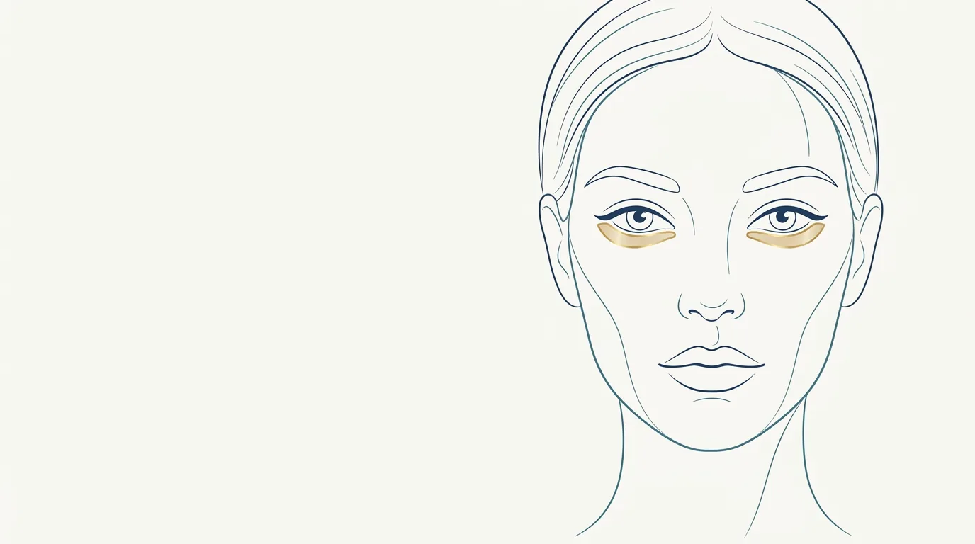
The lower eyelid runs from the margin of the lower lashes to the junction with the cheek. It is a narrow, delicate strip, but it is what determines whether the gaze looks rested or tired. In orofacial harmonization, the interest lies in the eyelid-cheek transition: when this transition forms a step or a groove, the area looks marked and ages the midface.
This page complements the one on dark circles, which covers the tear trough, and the overview of the periorbital region.
What are the anatomical boundaries of the lower eyelid?
In clinical practice, the lower eyelid is divided into two parts:
- Anterior lamella: skin and orbicularis oculi muscle.
- Posterior lamella: tarsus and conjunctiva.
Between them lies the orbital septum, which holds the orbital fat pads (medial, central, and lateral). Below, the orbital retaining ligament tethers the tissues to the rim and forms the palpebromalar groove laterally. At the inner corner, the tear trough ligament forms the tear trough.
With aging, three things tend to happen at the same time: the orbital fat may protrude, midface support decreases, and the skin thins. The result is the "step" between eyelid and cheek.
Vessels and nerves to watch
- Angular artery and branches of the ophthalmic artery at the medial corner.
- Infraorbital artery and nerve, exiting the foramen below the rim.
- Zygomaticofacial artery and lateral branches.
- Veins of the area, with drainage linked to the orbital circulation.
The lower eyelid is part of the periorbital area and should be treated as a high vascular-risk zone when filler is used.
What do patients usually ask for?
Concerns show up like this:
- "I have a step under my eye."
- "It looks like I have bags, even though I don't."
- "The area under my eye looks hollow."
- "I look tired in photos with overhead light."
The assessment needs to separate a step caused by loss of support (which may respond to filler) from a true fat pad and skin laxity (which generally do not respond and may be a surgical indication). Ask the patient to look up: the fat pad usually becomes more obvious, which helps tell them apart.
Which procedures can be done on the lower eyelid?
The most commonly used in orofacial harmonization are:
- Lower eyelid smoothing, with low G' hyaluronic acid to soften the eyelid-cheek transition.
- Under-eye filler, when the medial groove is the main component.
- Malar filler, to restore midface support and reduce the step with less product near the eye.
Technique, plan, and the choice between cannula and needle are at the discretion of the licensed professional, according to the rules of their professional council in Brazil.
What are the usual volumes for the lower eyelid?
Usual ranges in ml of hyaluronic acid, at the discretion of the licensed professional, after assessment:
| Situation | Usual range per side | Note |
|---|---|---|
| Subtle step | 0.2 to 0.5 ml | Often enough |
| Moderate step | 0.5 to 1 ml | Assess malar support together |
| Step with an obvious fat pad | Assess the indication | Filling may worsen the look |
| Durability | Generally 9 to 18 months | Area with little mobility |
Small volumes, with a follow-up to assess, are usually preferable to a complete correction in a single session.
Care, contraindications, and complications
General contraindications: active infection, pregnancy and breastfeeding, allergy to the components, and unrealistic expectations. In the lower eyelid, also assess prominent fat pads, significant laxity, chronic eyelid swelling, active eye allergies, and recent eyelid surgery.
Reported complications:
- Prolonged swelling, very characteristic of the area.
- Tyndall effect when the product sits superficially.
- Bruising, irregularities, and lumps.
- Worsening of the bag appearance, when filler was not the right indication.
- Vascular occlusion, with a risk of necrosis and visual complications.
Hyaluronidase must be available, with a vascular occlusion and visual complication protocol ready. See the overview of safety and complications.
How to assess the eyelid-cheek transition?
Assessing the lower eyelid calls for attention to details that change the indication:
- Looking up: a true fat pad usually becomes more obvious when the patient looks up.
- Skin pinch test: gives an idea of the laxity and elasticity of the eyelid.
- Overhead light: simulates everyday lighting and shows the shadow of the step.
- Comparing times of day: patients with significant morning swelling tend to respond worse to filler.
- Malar support: if the cheek has descended, the transition gets longer and the step more marked.
When fat pads and laxity predominate, the honest conversation is about referral, not volume. Recording these observations in a standardized photo helps explain them to the patient. See the guidance on standardized clinical photography.
How does the lower eyelid appear in Harmy?
The lower eyelid has no separate point in Harmy's marking step. It is represented by the Tear Trough point, which covers the tear trough and the lower eyelid transition. In the app's catalog, lower eyelid smoothing is linked to this same point.
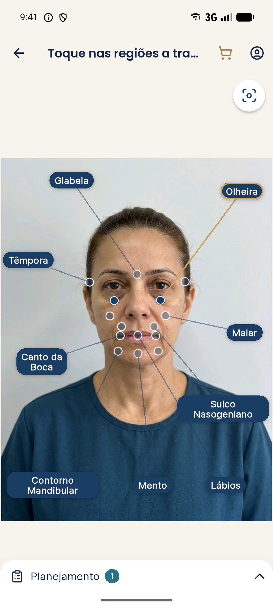
After marking Tear Trough, set the volume in the Plan (0.5 ml steps, from 0.5 to 5 ml). Because it is a bilateral area, the value is the total, split between both sides. The projection changes only the marked area, without retouching the skin, without makeup, and without changing the expression. See the step by step in marking areas and volumes.
How to talk to the patient about the lower eyelid
Patients with a lower eyelid concern often think they have a "bag" when they have a step, or the other way around. Showing a subtle illustrative simulation on their own photo helps explain what filler can soften and what depends on another approach.
Make clear that the projection is an illustration, and record the alternatives discussed in the informed consent. To handle frequent doubts, see patient objections and answers.
Frequently asked questions
What is the difference between the tear trough and the lower eyelid?
The tear trough, in the filler sense, is the groove at the inner corner. The lower eyelid covers the whole strip below the eyes up to the transition to the cheek, including the lateral part.
Does lower eyelid filler get rid of eye bags?
No. A true fat pad is not treated with filler and can even become more obvious. Filler can soften the step between the bag and the cheek in selected cases.
Is the lower eyelid area risky?
Yes. It is part of the periorbital area, with vessels connected to the eye's circulation and very thin skin. It requires experience, small volumes, and hyaluronidase on hand.
How do I plan the lower eyelid in Harmy?
The area has no separate point in the app. It is represented by the Tear Trough point, which covers the tear trough and the lower eyelid transition.
Harmy images are illustrative simulations generated by AI from the patient's photo to support the conversation between practitioner and patient. They are not a promise of results. Indication, technique, product and volume are solely the decision of the licensed practitioner. This content is for information only and does not replace a clinical assessment.