
Pigmentary, Vascular or Structural Dark Circles: How to Assess

"Dark circles" is a patient term, not a diagnosis. Behind it can be skin color (pigmentary dark circles), visible vessels (vascular dark circles), the shadow of a depression (structural dark circles), a fat bag or swelling, and almost always a mix of them. Classification is what defines whether filler makes sense, whether another approach comes first or whether the best choice is not to intervene.
This guide covers classification and indication. The anatomy of the tear trough, usual volumes and filler complications are on the area page dark circles and in the procedure under-eye filler.
What are the types of dark circles?
The table summarizes the types most cited in clinical assessment, how to recognize them and what tends to help. Indication, technique and product choice are at the discretion of the qualified professional, according to the rules of their professional council.
| Type | Main cause | How it looks | Response to filler | What tends to help |
|---|---|---|---|---|
| Pigmentary | Excess melanin in the eyelid skin | Brownish tone, sometimes extending to the upper eyelid | Does not treat the color | Skin care, sun protection, dermatologic treatments |
| Vascular | Thin skin that shows vessels and muscle | Bluish, purple or pinkish tone, worse with tiredness | Limited response | Skin quality, habits, individual assessment |
| Structural | Tear trough depression and loss of midface support | Shadow that changes with light | Usually responds well | Tear trough filler and cheekbone support |
| Due to a fat bag | Herniation of the orbital fat pads | Volume above the groove, worse when looking up | Filling may worsen it | Assessment of alternatives, including surgical ones |
| Due to swelling | Fluid retention in the eyelid | Puffiness that varies through the day, worse in the morning | Relative contraindication | Investigate causes before any injectable |
| Mixed | Combination of the above | Color and shadow at the same time | Partial improvement | Treat the components in stages |
The mixed type is the most common in practice. The role of assessment is to say how much of the complaint comes from each component, because that defines what the patient can expect.
How do you tell the types apart at the visit?
A few simple tests, done with the patient seated and good lighting, help separate color from shadow.
Light test
Observe the area with frontal light and then with light from above, like ceiling light. A structural dark circle is shadow: it worsens with overhead light and improves a lot with frontal light or when the patient tilts the face slightly upward. Pigmentary and vascular color remain there, whatever the light.
Compression test
Gently press the skin with a clear glass slide or the pad of a finger. The vascular component tends to blanch under pressure and return on release. The pigmentary component does not change. Some professionals use a Wood's lamp, which accentuates more superficial pigment.
Looking up and to the sides
Ask the patient to look up. Orbital fat bags usually become more obvious in that position, and the volume above the groove increases. This finding changes the indication.
Variation through the day
Ask whether the dark circle is worse in the morning, or whether it varies with sleep, salt, alcohol or allergy flare-ups. Large variation suggests swelling, and frequent eyelid swelling calls for caution with any injectable in the area.
What should you ask in the medical history?
History helps as much as the exam:
- Since when? Dark circles since childhood, with a family history, suggest a constitutional, pigmentary or vascular component.
- Allergies and rhinitis? Chronic congestion is associated with dark circles and swelling.
- Sleep, smoking and sun exposure? They influence skin color and quality.
- Eye drops in use? Some glaucoma eye drops are associated with darkening and fat loss around the eyes.
- Recent weight loss? May accentuate the structural component.
- Previous procedures in the area? Prior filler, eyelid surgery or laser change the reading and the risk.
These points belong in the medical history in HOF and in the patient record.
When is filler indicated?
The classic indication is a predominantly structural dark circle: depression of the tear trough, a marked transition between eyelid and cheek and shadow that changes with light. The skin needs reasonable thickness and there should be no obvious bag or frequent swelling.
Even in these cases, many professionals first assess midface support. Restoring support with cheekbone filler can improve the dark circle with less product in the tear trough, which is an area of thin skin and high vascular risk. The transition with the eyelid is discussed in lower eyelid smoothing.
When not to fill, or not to fill yet?
- Obvious fat bag, which tends to look larger afterward.
- Frequent or chronic eyelid swelling.
- Very thin, translucent skin, with a higher risk of the Tyndall effect.
- Complaint predominantly about color, pigmentary or vascular.
- Expectation that the dark circle will "disappear" or lighten.
Does under-eye filler remove the purple color?
It is the most common question, and the honest answer is: generally, no. Filler corrects the depression and reduces the shadow. If the purple comes from visible vessels through thin skin, it stays. In some patients, when the shadow decreases, the whole area looks lighter, and this can give the impression that the color improved; the skin color itself does not change.
For the skin quality component, some plans include approaches such as the skin booster and, more recently, polynucleotides, always checking the registration and the approved indication of the product.
Why does under-eye filler sometimes turn purple?
Two pictures explain most complaints, and they are very different:
| Picture | When it appears | How it evolves | What is usually done |
|---|---|---|---|
| Bruise | Right after or the next day | Changes color (purple, green, yellow) and disappears in one to two weeks | Guidance and follow-up |
| Tyndall effect | Visible after the swelling goes down | Stable bluish tone, sometimes with relief, that does not improve on its own | Assessment and, generally, hyaluronidase, at the professional's discretion |
Explaining this difference before the procedure avoids anxiety: the patient knows that a purple that changes color is expected, and that a bluish tone that does not change should be shown to the professional.
How do you explain the classification to the patient?
Patients usually arrive expecting "the dark circles to disappear". Classification lets you swap that expectation for a concrete explanation: what is shadow, what is color, what each treatment achieves.
How do dark circles appear in Harmy?
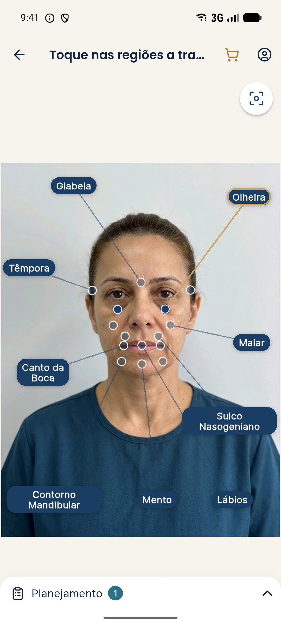
In the Plan, volume is set in ml (0.5 ml step, from 0.5 to 5 ml); for a bilateral area, the value is the total, split between the two sides. Remember that the projection is an illustrative simulation, not a promise of results.
Dark circle assessment checklist
- Light test done and photographed (frontal and overhead).
- Compression test to separate vascular from pigmentary color.
- Patient observed looking up, to assess bags.
- History of swelling, allergy, eye drops and previous procedures recorded.
- Predominant component defined and explained to the patient.
- Cheekbone support assessed before the tear trough.
- Expectation about color agreed in writing.
- Hyaluronidase and visual complication protocol ready, if filler is used.
Frequently asked questions
Does under-eye filler remove the purple color?
Generally, no. Filler treats the depression of the tear trough, that is, the shadow. The purple color of vascular origin and pigmentary darkening of the skin do not change with the product, although the whole area may look lighter when the shadow decreases.
Why does under-eye filler turn purple?
In the first days, the purple is usually a bruise, which changes color and disappears within one to two weeks. A stable bluish tone that does not improve suggests the Tyndall effect, linked to product in a superficial plane, and should be assessed by the professional.
How do I tell whether a dark circle is pigmentary or vascular?
A pigmentary dark circle has a brownish tone and does not lighten when the skin is pressed. A vascular one is bluish or purple, usually gets paler under compression and changes with tiredness and sleep. Many patients have both components.
Can someone with a bag under the eye have under-eye filler?
An obvious fat bag is usually a reason not to fill the tear trough, because the volume above the groove tends to look larger afterward. In these cases, assessing other approaches, including surgical ones, comes first.
Harmy images are illustrative simulations generated by AI from the patient's photo to support the conversation between practitioner and patient. They are not a promise of results. Indication, technique, product and volume are solely the decision of the licensed practitioner. This content is for information only and does not replace a clinical assessment.