
Facial Bone Resorption With Age: Maxilla, Mandible, and Orbit
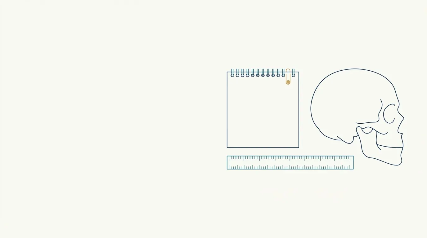
- Highlighted area
Quick facts
- Core idea
- The facial skeleton changes with age: the orbit widens, the maxilla recedes, and the mandible loses height and projection.
- Consequence
- Soft tissue loses support, which contributes to hollow under-eyes, folds, descent of the midface, and loss of the jawline.
- Clinical implication
- Treating only skin or fat may not be enough when the problem has a bony component, and the plan should consider the structure.
- Limit
- The speed and extent of resorption vary between people, and this text describes tendencies, not individual rules.
When you look at an aging face, what first draws attention is usually the skin and the lines. Yet part of what looks like "sagging" or "lack of volume" has a deeper origin: the facial skeleton itself changes. The orbit enlarges, the maxilla recedes, the mandible loses height. The soft tissues, which rest on that support, follow the change. Understanding the role of bone helps the orofacial harmonization (HOF) professional explain to the patient why certain concerns come back, why some areas call for more care, and why the treatment plan should consider the structure. This guide describes the tendencies at a review level. Indication, product, and technique are the decision of the qualified professional, according to the product label and the rules of their professional council.
Does facial bone really change?
Yes. Imaging and anatomy studies describe remodeling of the facial skeleton throughout adult life. The speed and intensity vary, but the trend is consistent in some regions. Bone is not a static structure: it is living tissue that remodels in response to hormones, mechanical load, nutrition, and disease.
In practice, the result is a loss of support for the layers above it: muscles, fat, and skin. This loss adds to fat atrophy, loosening of the ligaments, and loss of skin elasticity. See the overview in facial aging by layers.
What happens in each region?
| Region | Described tendency | Apparent effect on soft tissue |
|---|---|---|
| Orbital cavity | Increase in diameter, with recession of the orbital rim, especially inferior and lateral | Deeper hollow under the eye, more visible bags, shadow under the eye |
| Maxilla | Retrusion and loss of projection, with remodeling of the angle and of the premaxilla region | Loss of cheek support, deepening of folds, change in the base of the nose and the upper lip |
| Mandible | Reduced height of the body and chin, change in the mandibular angle, alveolar bone resorption with tooth loss | Loss of contour definition, formation of jowls, change in the lower third |
| Piriform aperture (nose) | Widening | Change in projection and support of the nasal tip |
| Frontal and temporal region | More subtle remodeling, with change in contour | Apparent loss of volume at the temples |
These patterns are described tendencies and vary between sexes and between individuals. For specific reasoning, see periorbital anatomy for injectables, aging of the midface, and aging of the chin and jaw.
How does this explain common concerns?
Some concerns seen in the clinic make sense when you think about the skeleton:
- Under-eye hollow that "won't go away with anything." When there is recession of the orbital rim and widening of the orbit, the shadow under the eye has a structural basis, and surface treatments have limited effect.
- Cheek that "dropped" and a fold that deepened. A retruded maxilla offers less support, which changes the support vector of the midface.
- Jawline contour that disappeared. Loss of bone height and projection, added to the descent of fat, forms jowls and undoes the clean line.
- Face that "shrank." Reduction of the bony base leaves excess soft tissue, giving the appearance of sagging.
- Profile that changed. Changes in the maxilla and chin alter the projection of the lip and nose. See profile changes with age.
Which factors influence resorption?
Some of these items point to other areas of health. When bone disease is suspected, referral to the appropriate physician is worth more than any aesthetic procedure. See the relationship with the dental setting in HOF integrated with dentistry.
How does planning change when bone is taken into account?
Thinking about structure changes the logic of treatment. Instead of treating only the fold, the hollow under the eye, or the jowl, the professional asks which layer needs support. Some usual lines of reasoning:
- Restore support where it was lost, at strategic projection points, before treating what lies above. In some cases this can reduce the need for volume in areas with less support.
- Avoid compensating for structural loss with excess superficial volume, which leads to a heavy look. See pillow face.
- Consider products with characteristics suited to the function, such as greater lifting capacity in projection areas. See G prime and calcium hydroxylapatite in the jawline.
- Use threads, biostimulators, or other strategies depending on the layer and the indication. See biostimulators: overview.
- Recognize the limit of the technique: when structural change is significant, surgery or an orthognathic approach may be part of the conversation.
- Involve dentistry, especially in cases of tooth loss and occlusion problems.
How do you communicate this to the patient?
Explaining the bony base helps align expectations, but it calls for care so as not to frighten. Patients usually think the face ages because of "sagging skin." Showing that there is a structure underneath that has changed helps justify why one treatment alone may not be enough.
Use accessible language and avoid alarming terms ("collapsing face"). Focus on what can be improved.
How does time shape this change?
This timeline is a simplification. The age at which each phase appears varies widely.
Which myths should you avoid?
What should you document?
In the patient record, record the layer-by-layer assessment (skin, fat, ligaments, bone), the profile and three-quarter observations, the patient's concerns, relevant dental and hormonal history, standardized photos, the plan, and the expectations discussion. If there was a referral to dentistry or another specialty, note it. Standardized, comparable photos are the best reference for following a slow process. See standardized clinical photography.
Where does Harmy fit in?
Harmy starts from a front-facing photo and simulates volume in marked areas. In areas such as Cheekbone, Tear Trough, and Jawline, the simulation can help show the patient the effect of adding support where there is structural loss.
Because the app does not assess bone structure, clinical assessment remains irreplaceable.
In summary
Facial aging includes the skeleton. The orbit, maxilla, and mandible remodel, and soft tissues lose support. This knowledge helps you understand concerns, choose where to add support, avoid excess, and align the conversation with the patient. Layer-by-layer assessment, attention to factors such as teeth and hormones, and respect for the limits of each technique make the difference between treating the symptom and planning based on structure.
Frequently asked questions
Does the bone of the face really change with age?
Yes. The anatomy literature describes remodeling of the facial skeleton throughout life, with widening of the orbit, retrusion of the maxilla, and loss of height and projection in the mandible, varying between individuals and between sexes.
Does bone resorption explain under-eye hollows?
It can contribute, because widening of the orbit and recession of the rim change the support of the lower eyelid and the cheek. Other factors, such as fat, skin, and pigmentation, also play a part.
Does filler over bone restore what was lost?
It can compensate for part of the loss of projection in strategic areas, at the professional's discretion. It does not reverse resorption and does not replace plans that call for other approaches.
Is bone resorption the same in everyone?
No. Age, sex, genetics, dental status, hormones, habits, and diseases influence the pattern. That is why planning starts from individual assessment and not from a single model.
Does tooth loss speed up resorption of the mandible?
Tooth loss is associated with resorption of the alveolar bone, which affects the lower third of the face. That is why working together with dentistry makes a difference in planning.
Harmy images are illustrative simulations generated by AI from the patient's photo to support the conversation between practitioner and patient. They are not a promise of results. Indication, technique, product and volume are solely the decision of the licensed practitioner. This content is for information only and does not replace a clinical assessment.