
Hyaluronic acid in the preauricular area: what to assess
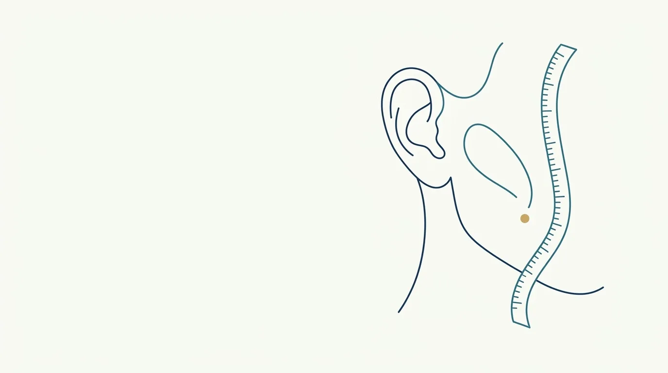
- Highlighted area
Quick facts
- Area
- The strip in front of the ear, between the zygomatic arch and the angle of the jaw.
- Neighboring structures
- Parotid gland, superficial temporal artery, transverse facial artery and branches of the facial nerve.
- Common concern
- Flattening or loss of convexity of the lateral facial contour, a face that looks unsupported.
- Central caution
- An area of superficial vessels and nerves, with little room for improvisation.
The preauricular area is the strip of the face immediately in front of the ear. It makes the transition between the temple, the zygomatic arch and the angle of the jaw, and when it loses support, the lateral contour of the face looks flattened or "sagging". It is an area increasingly discussed in rejuvenation plans for the midface and lateral face, but it concentrates structures that do not forgive improvisation.
This page does not teach technique. It lays out the anatomy of risk, what to assess before thinking about hyaluronic acid, the alternatives and what to document. Product, volume and approach are decisions for the licensed professional, according to the rules of their professional council in Brazil.
Which structures cross the area?
The area is small and dense. Review before any plan:
- Parotid gland: a large salivary gland in front of and below the ear. It is covered by a fascia, and the facial nerve passes through it. Material placed deeply can come close to it.
- Facial nerve: its temporal and zygomatic branches leave the parotid and spread across the face. The temporal branch and the zygomatic branch are superficial in some places, which increases vulnerability.
- Superficial temporal artery: a terminal branch of the external carotid, it rises in front of the ear and is palpable in many patients.
- Transverse facial artery: runs through the space between the zygomatic arch and the parotid.
- Zygomatic arch and retaining ligaments: limit the space and anchor the skin.
For the map of layers, see layers of the face and facial aging by layers.
What happens to the lateral contour with age?
Aging of the lateral third is not only fat loss. There is bone resorption of the zygomatic arch and lateral skeleton, thinning of the fasciae, descent of fat compartments and laxity of the ligaments. The result, seen from the front, is a face that loses its lateral "curve" and looks narrow or hollow at ear level.
This picture resembles that of the temple, so the assessment should cover both. See aging of the temples and temporal anatomy for injectables.
| Finding | May indicate | General direction |
|---|---|---|
| Hollow temporal fossa and prominent zygomatic arch | Temporal and bone volume loss | Assess the temple as a unit |
| Straight lateral contour, no curve, loose skin | SMAS and ligament laxity | Consider support strategies; gel alone may not be enough |
| Shadow in front of the ear with a full cheek | Compartment descent | Assess the midface as a whole |
| Parotid ptosis or glandular volume | Anatomical variation | Caution: gel does not correct it and may worsen it |
When does it make sense to discuss gel?
In the practical literature and in courses, filling the lateral area appears as part of a support plan, usually combined with assessment of the cheekbone and temple. The discussion is more frequent in middle-aged and older patients with loss of lateral convexity and less relevant in young patients.
Before recommending, it is worth asking whether the expected benefit justifies the risk, whether the patient has realistic expectations and whether there are alternatives with a better safety profile for their case, such as biostimulators used by trained professionals (see biostimulator in the preauricular area), threads or other support approaches.
What are the risks and how to recognize them?
The adverse events described for this area fall into categories:
- Vascular: occlusion of branches of the superficial temporal artery or of small skin vessels, with pain, blanching or a purplish color. Read vascular occlusion in the temples as a management reference for a neighboring area.
- Nerve: weakness of a facial nerve branch, which may show as asymmetry when smiling or frowning.
- Glandular: parotid swelling or pain, rare.
- Aesthetic: palpable nodule, irregularity, a "puffy" look and asymmetry.
- Bruising: both superficial and deep, with possible persistent swelling.
What are the alternatives to gel in this area?
Gel is one tool, not the only one. Depending on the cause:
- Collagen biostimulator, applied by a trained professional, for gradual tissue support, with its own risks;
- Thread lifts, discussed in PDO threads in the midface, with their own indications and complications;
- Skin and energy-based treatments, such as radiofrequency, when the problem is superficial laxity;
- An approach to the midface as a whole, instead of treating only the preauricular strip.
Each option has limits and risks. The central point is that the plan should come from diagnosing the cause.
Assessment checklist
How does it evolve after the procedure?
More in post-procedure instructions and clinical follow-up visit.
What the app can (and cannot) show
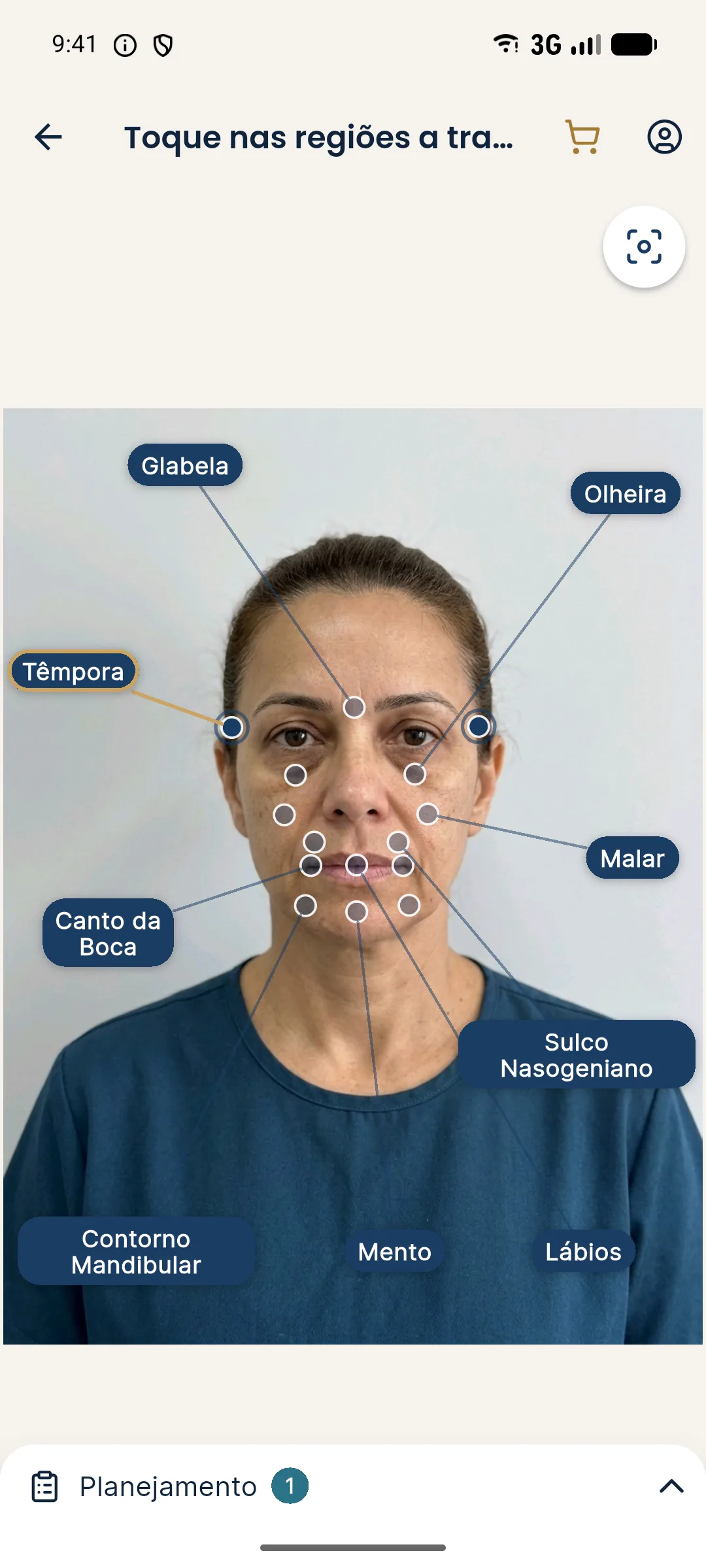
Harmy marks the Temple and Cheekbone areas, which sit beside this strip. The projection helps show the patient the effect of a support plan in the temple and cheek, using only the frontal photo. There is no "preauricular" point in the app, and the projection does not represent a profile or 45-degree view.
Myth or fact
Summary for practice
Before touching the preauricular area, define whether the complaint is about volume, support or gland, review the anatomy of vessels and nerves, offer alternatives and record specific consent. If doubt persists, treating the neighboring areas or referring to a colleague with more experience in this area is a legitimate professional decision. See also hyaluronic acid by area, temple filler and vascular danger zones of the face.
Frequently asked questions
What is the preauricular area?
It is the area in front of the ear, between the temple and the jaw, where the parotid gland, vessels such as the superficial temporal artery and branches of the facial nerve are found. It forms the lateral contour of the face.
Can the preauricular area be filled with hyaluronic acid?
It is described in some harmonization plans, but it is an area of complex anatomy and many professionals approach it with caution or prefer other strategies. The decision rests with the licensed professional, after assessment.
What are the risks of preauricular filler?
Bruising, swelling, nodule, asymmetry, injury to a nerve branch, vascular compromise and, by proximity, damage to the parotid gland. Hyaluronidase should be available.
What is the difference between filling the preauricular area and the temple?
The areas connect, but have different anatomy: the temple has fasciae and deep temporal vessels, while the preauricular area involves the parotid, the superficial temporal artery and the facial nerve. Each needs its own reading.
Harmy images are illustrative simulations generated by AI from the patient's photo to support the conversation between practitioner and patient. They are not a promise of results. Indication, technique, product and volume are solely the decision of the licensed practitioner. This content is for information only and does not replace a clinical assessment.