
Temporal Region Anatomy for Injectables
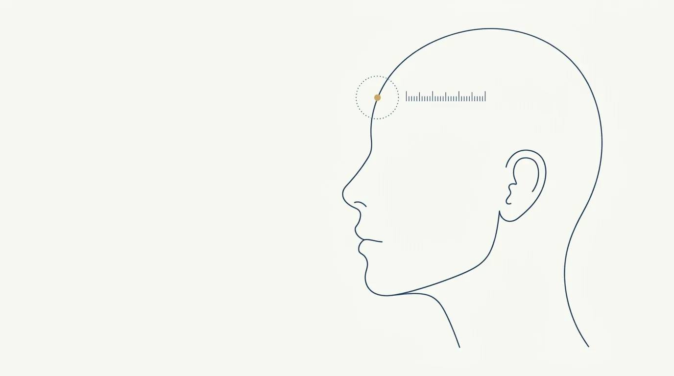
- Nerve
- Highlighted area
Quick facts
- Structure
- Skin, subcutaneous tissue, temporal fasciae, fat pad and temporalis muscle over the bone of the temporal fossa.
- Vessels of concern
- Superficial temporal, middle temporal and middle temporal vein (sentinel vein), plus deep branches close to the muscle.
- Nerve of concern
- Temporal branch of the facial nerve, which runs in a superficial layer in the region.
- Main risk
- Vascular occlusion and hematoma, with possible damage to the skin or, more rarely, to the eye's circulation, plus irregularity.
The temple is an area that changes the face without the patient realizing why. When it hollows, the forehead looks narrower, the brow ridge heavier and the eyes more tired. Anyone assessing this area needs to understand that it is not an "upper cheek": it is a compartment with well-defined layers, vessels of significant caliber and a superficial nerve. This guide reviews the anatomy, what changes with age and how that translates into indication and risk. Technique, injection plan and volumes are decisions for the qualified professional, following the label and the rules of their professional council.
How is the temporal region organized?
The temporal fossa is a bony depression on the side of the skull, bounded by the zygomatic arch, the temporal line and the ridge of the frontal bone. It is filled with layers, from the surface to the bone:
- Skin, relatively thin and mobile.
- Subcutaneous tissue, with fat and vessels.
- Temporoparietal fascia, a layer that contains the superficial temporal artery and the temporal branch of the facial nerve.
- Temporal fascia, which covers the muscle and splits into superficial and deep layers in its lower part.
- Temporal fat pad, between the layers of the fascia.
- Temporalis muscle, a fan-shaped muscle of mastication.
- Periosteum and bone, at the bottom of the fossa.
The exact naming of the layers varies between authors, but the idea is fixed: there are several stacked layers, with critical structures at different levels. The big picture is in layers of the face and in facial aging by layers.
What changes over time
Temporal hollowing is multifactorial. In general, these add up:
- Bone resorption of the fossa and the orbital contour;
- Loss of the temporal fat pad;
- Atrophy of the temporalis muscle, especially in patients with weight loss, no bruxism or advanced age;
- Loss of skin elasticity, which reveals the bony relief.
Other contexts also cause temporal concavity: marked weight loss, wasting diseases and the use of certain medications. See the picture in facial volume loss after weight loss. A muscle enlarged by bruxism, on the other hand, can produce a different look with its own treatment, as covered in botulinum toxin for bruxism.
Vessels of concern
The temporal region is supplied by branches of the external carotid artery and has connections with neighboring territories.
| Vessel | Where it usually lies | Why it matters |
|---|---|---|
| Superficial temporal artery | Superficial layer, with frontal and parietal branches | Significant caliber; course visible or palpable in some patients |
| Middle temporal artery | Pierces the fascia and runs in a deeper layer | Arises from the superficial artery; connections with other branches |
| Deep temporal arteries | Accompany the temporalis muscle | Supply muscle and bone; possible bleeding in accidents |
| Middle temporal vein | Pierces the fascia and is visible in some patients | Anatomical landmark (sentinel vein), but inconsistent |
These structures have anastomoses with branches that go to the face, the orbit and the scalp. The general principle of the connections is in vascular anastomoses of the face. For the safety reasoning, read vascular danger zones of the face.
Nerves and sensation
Two branches deserve mention:
- Temporal branch of the facial nerve: supplies the frontalis muscle and the upper part of the orbicularis oculi. Running in a superficial layer, it is vulnerable to trauma. An injury can cause asymmetry of the brow and forehead.
- Zygomaticotemporal nerve: sensory, it supplies the skin of the lateral temple. Changes in sensation can appear after procedures.
If the patient reports tingling, burning or loss of sensation afterward, record it, examine and follow up. It may be benign and temporary, and it may need evaluation.
How anatomy guides the indication
With the layers in mind, the assessment becomes more objective.
In many cases the temple is part of a broader plan that includes the cheekbone, the brow and the forehead. See also biostimulator in the temples as an alternative in certain situations.
How to explain it to the patient
Records and consent
Document the exam, the photos, the classification of the concavity, the product, the lot number, the instructions and the follow-up. The consent should name hematoma, asymmetry, nerve injury and vascular complication. The record structure is in patient records and documentation in HOF.
Where Harmy comes in
Common mistakes in temporal assessment
- Looking only from the front. Temporal concavity shows best from a three-quarter view and from above, with side light.
- Forgetting bruxism. An enlarged muscle changes the contour and can explain different complaints.
- Confusing bone, muscle and fat. Each component responds differently, and the plan changes.
- Underestimating weight lost. Recent weight loss can hollow the temple quickly, and the picture does not always resolve with an injectable.
- Not palpating before deciding. Veins, pulses and bony irregularities guide caution.
Questions patients often ask
- "Why did my temple hollow?" Explain that bone, fat and muscle change over time and that together they alter the contour.
- "Does this make my face look older?" Say that the temple influences how the forehead and eyes read, without promises about appearance.
- "Is it dangerous?" Be honest: it is an area with important vessels and nerves, which is why being prepared for emergencies is part of the procedure.
- "How long does it last?" Give a range and make clear it varies with the product and the patient.
Finally, it is worth recording the history of the concern: since when the temple has looked more hollow, whether there was weight loss, a change in medication, illness or recent treatment. This information separates gradual aging hollowing from conditions that deserve clinical investigation before any injectable, and helps align the patient's expectations with what the procedure can offer.
Frequently asked questions
Why do the temples hollow with age?
Because of bone resorption, loss of the fat pad and atrophy of the temporalis muscle, which together reduce the volume of the temporal fossa. The result is a concave temple, which changes the contour of the forehead and the eyes.
Is the temple a high-risk area?
Yes. There are vessels of significant caliber, with individual variation, and a superficial branch of the facial nerve. Hyaluronidase on hand, anatomical knowledge and an emergency protocol are essential.
What is the sentinel vein?
It is a middle temporal vein that pierces the fascia and may show under the skin of the temple. It is described as an anatomical landmark, but variations are common, and its presence does not rule out other risks.
Does every patient with concave temples need filler?
No. The hollow may be constitutional or mild, and the indication depends on the concern, the face as a whole and the professional's assessment. In some cases, the right approach is to monitor.
Harmy images are illustrative simulations generated by AI from the patient's photo to support the conversation between practitioner and patient. They are not a promise of results. Indication, technique, product and volume are solely the decision of the licensed practitioner. This content is for information only and does not replace a clinical assessment.