
Anteromedial Cheek Area: Hyaluronic Acid Indication
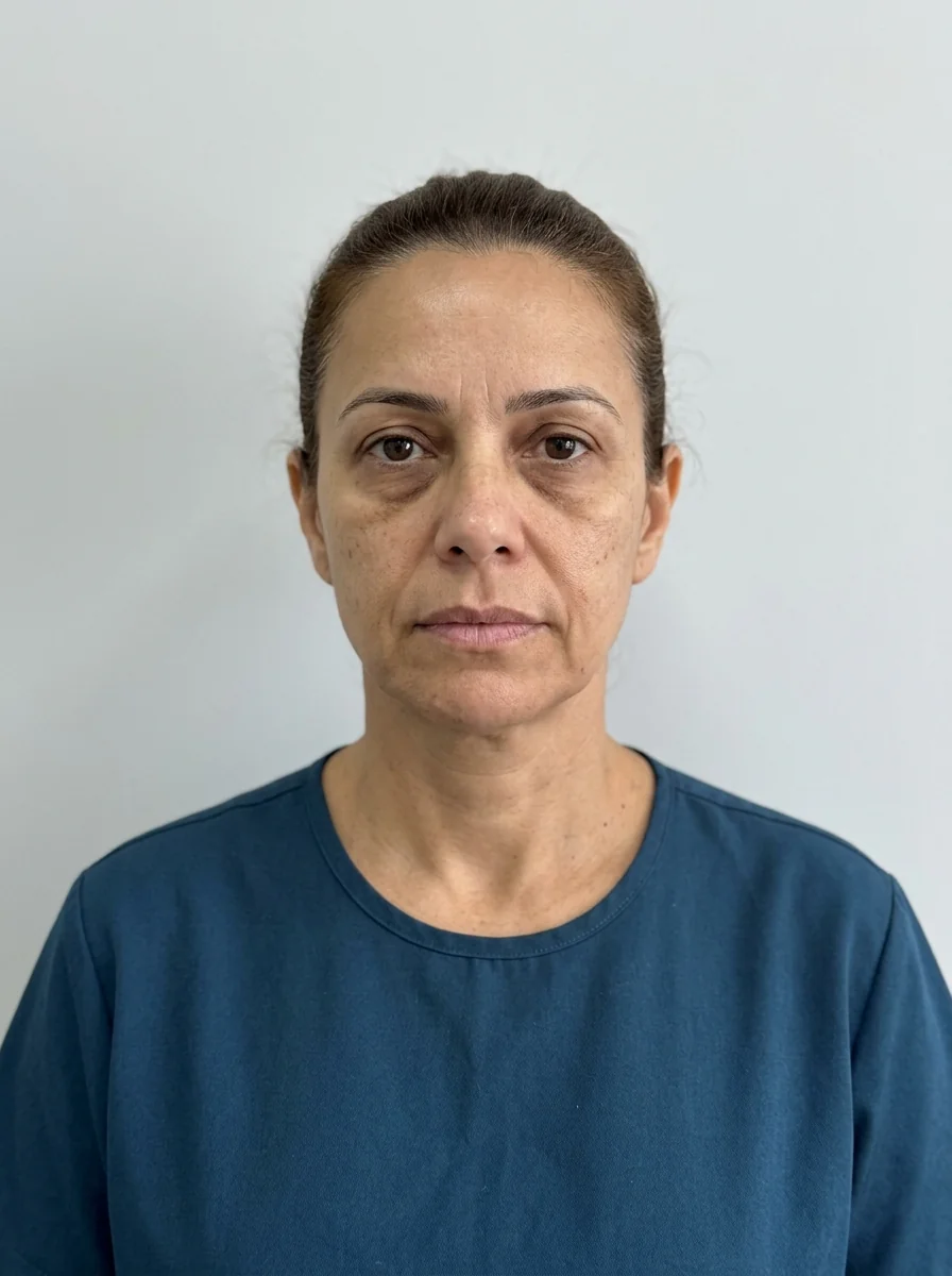
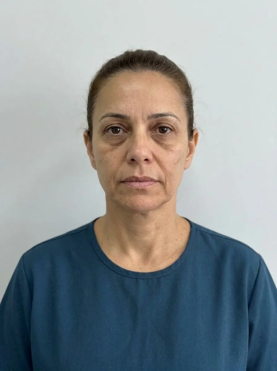 BeforeSimulation
- Nerve
- Highlighted area
Quick facts
- Area
- Anteromedial cheek, next to the palpebromalar and mid-cheek grooves
- Structures of concern
- Zygomatic ligament, angular artery and infraorbital nerve
- Usual concern
- Loss of midface support and a marked transition to the lower eyelid
- Risks
- Swelling, irregularity, Tyndall effect and vascular occlusion
- Prerequisite
- Assessment of the midface and hyaluronidase available
The anteromedial cheek is where the cheek meets the nose and the lower eyelid. It is also where several concerns overlap: under-eye circles, a marked palpebromalar groove, a tired look, an "empty" midface. That is why it is tempting to treat it, and why it requires an assessment that separates what is loss of support from what is skin, eyelid fat or pigment.
This content is a review of anatomy, indication and risks for the orofacial harmonization (HOF) professional. It is not a step-by-step technique. Product, volume and approach are the decision of the licensed professional, after assessment, according to the rules of their professional board and the product's instructions for use.
What is in the anteromedial cheek area?
The area lies between three visible landmarks: the palpebromalar groove, the mid-cheek groove and the nasolabial fold. In depth, the design is one of fat compartments separated by retaining ligaments.
The zygomatic ligament anchors the skin and superficial tissue to the bone and defines part of the transition between the lower eyelid and the cheek. The prezygomatic space and the deep cheek fat (SOOF, see SOOF) take part in midface support. In practical terms: when deep support decreases, the skin and superficial fat descend, and the groove from the eyelid to the cheek shows more.
The overview by layers is in facial aging by layers and midface aging.
When is treatment usually considered?
Among the most cited situations:
- Loss of midface support, with a "tired face" look.
- A marked palpebromalar or mid-cheek groove, associated with loss of cheek volume.
- An accentuated transition between the lower eyelid and the cheek.
- Integrated planning of under-eye hollows, cheek and nasolabial fold.
Before talking about gel, assess the cause. An "under-eye circle" can be shadow from volume loss, pigment, visible vasculature or a fat pad, and each calls for a different strategy. The page under-eye circles: types and assessment organizes this separation, and malar bags: assessment helps avoid confusing it with swelling or a bag.
| What the patient describes | What may be behind it | Does filler solve it? |
|---|---|---|
| "Hollow under-eyes" | Loss of support and shadow | Sometimes, after assessment |
| "Dark under-eyes" | Pigment or vasculature | Generally not |
| "Bag under the eye" | Eyelid fat or swelling | May make it worse |
| "Tired face" | A combination of factors | Depends on the whole picture |
| "Sagging cheek" | Laxity and loss of support | In part |
What are the vascular and nerve risks?
The angular artery and the angular vein run near the inner corner of the eye and connect the facial territory with that of the orbit, which makes the medial area relevant from a vascular standpoint. The infraorbital nerve exits the bone through the infraorbital foramen and carries sensation to the upper lip and cheek. See also infraorbital vascular occlusion and periorbital anatomy for injectables.
The most discussed risks:
- Vascular occlusion, with local ischemia and, in rare cases, visual consequences.
- Swelling, more pronounced in the transition to the lower eyelid.
- Tyndall effect, when the gel is superficial in thin skin.
- Irregularity and nodule, mainly if the product is poorly suited to the area.
- Paresthesia, from contact with the infraorbital nerve (see paresthesia).
- Excess volume, which changes facial proportion and can give a "puffy cheek" look (see pillow face).
How to assess the patient?
A history of eyelid surgery is relevant, because the anatomy may be altered (see harmonization after blepharoplasty). Conditions such as Graves and Sjögren change the indication, as discussed in Graves ophthalmopathy and HOF and Sjögren and the periorbital area.
How to think of the cheek as part of the whole?
The anteromedial cheek is rarely an isolated problem. Its relationship with the lateral cheek, the zygoma and the nasolabial fold determines whether the face looks balanced. Reinforcing midface support can soften the transition to the nasolabial fold, but the opposite also happens: too much volume in the cheek can accentuate the fold if support is not balanced.
See the Cheekbone area, the cheek filler procedure and the page hyaluronic acid by area for the logic of product choice, which depends on the gel's projection capacity and hydrophilicity.
What does Harmy show and what does it not?
In Harmy, the Cheekbone point lets you set the ml and the intensity (Subtle, Moderate or Pronounced) and generate an illustrative projection from a front-facing photo. The app does not distinguish the anteromedial part from the lateral one; the projection shows the cheek area as a whole, which helps in talking about midface support. It is an illustrative simulation, not a promise of results. During the consultation, presentation mode (full screen with only the slider) and the magnifier on the treated area help show the difference without distraction.
What if something goes off plan?
For swelling, irregularity or a bluish tone, the approach depends on the picture, and hyaluronidase is an option when indicated. With signs of vascular occlusion (pain, pallor, livedo, visual change), follow the emergency protocol and refer without delay when there are visual symptoms. For preparation, see emergency kit for the HOF office. Follow-up and records are in clinical follow-up after HOF.
In summary
The anteromedial cheek is an area of high aesthetic value and demanding anatomy. The safest indication starts with a clear diagnosis of what is causing the tired look, continues with conservative volumes and ends with follow-up and records. Without a diagnosis, the best injection is the one that is not done.
Frequently asked questions
What is the anteromedial cheek area?
It is the part of the cheek closest to the nose and the lower eyelid, near the palpebromalar groove and the mid-cheek groove. It is usually assessed when looking at midface support and the transition to the infraorbital area.
Is filling the anteromedial cheek safe?
No procedure is risk-free. The area has relevant vessels and nerves, and injection calls for anatomical knowledge, conservative volume and hyaluronidase on hand.
Does anteromedial cheek filler help with dark circles?
In some cases the loss of cheek support contributes to the look of under-eye hollows, and the professional can assess the midface as a whole. Not every under-eye concern responds to filler, and that depends on the type.
How long does hyaluronic acid last in the cheek?
Duration varies with the product, the volume, the area and the patient, and is usually measured in months. The professional advises on follow-up depending on the case.
Harmy images are illustrative simulations generated by AI from the patient's photo to support the conversation between practitioner and patient. They are not a promise of results. Indication, technique, product and volume are solely the decision of the licensed practitioner. This content is for information only and does not replace a clinical assessment.