
Digital Visual Planning in Facial Harmonization: What It Is

Quick facts
- Definition
- Using photos and digital tools to record the assessment and visualize the plan before injecting
- Steps
- Standardized photo, assessment, marking, simulation, conversation, written plan
- Within scope
- Communication, aligning expectations, record keeping and quotes
- Outside scope
- Diagnosis, choice of technique and guaranteed results
Digital visual planning is the part of the visit where the plan stops existing only in the professional's head and starts existing in an image the patient can see. It is not a procedure or a technique. It is a way to organize the assessment, communicate and keep records. Used well, it reduces misunderstandings; used badly, it becomes a sales tool.
Here is what it is, the steps it breaks into, what fits inside it and what must stay outside.
What goes into digital visual planning?
Five elements make up the planning, and you can adopt just some of them:
- Standardized photography. Frontal, profile and oblique, with controlled light, distance and expression. Without this, any later comparison loses value. See standardized clinical photography.
- Recorded facial assessment. Thirds, symmetry, proportions and concerns noted, preferably on a plan sheet.
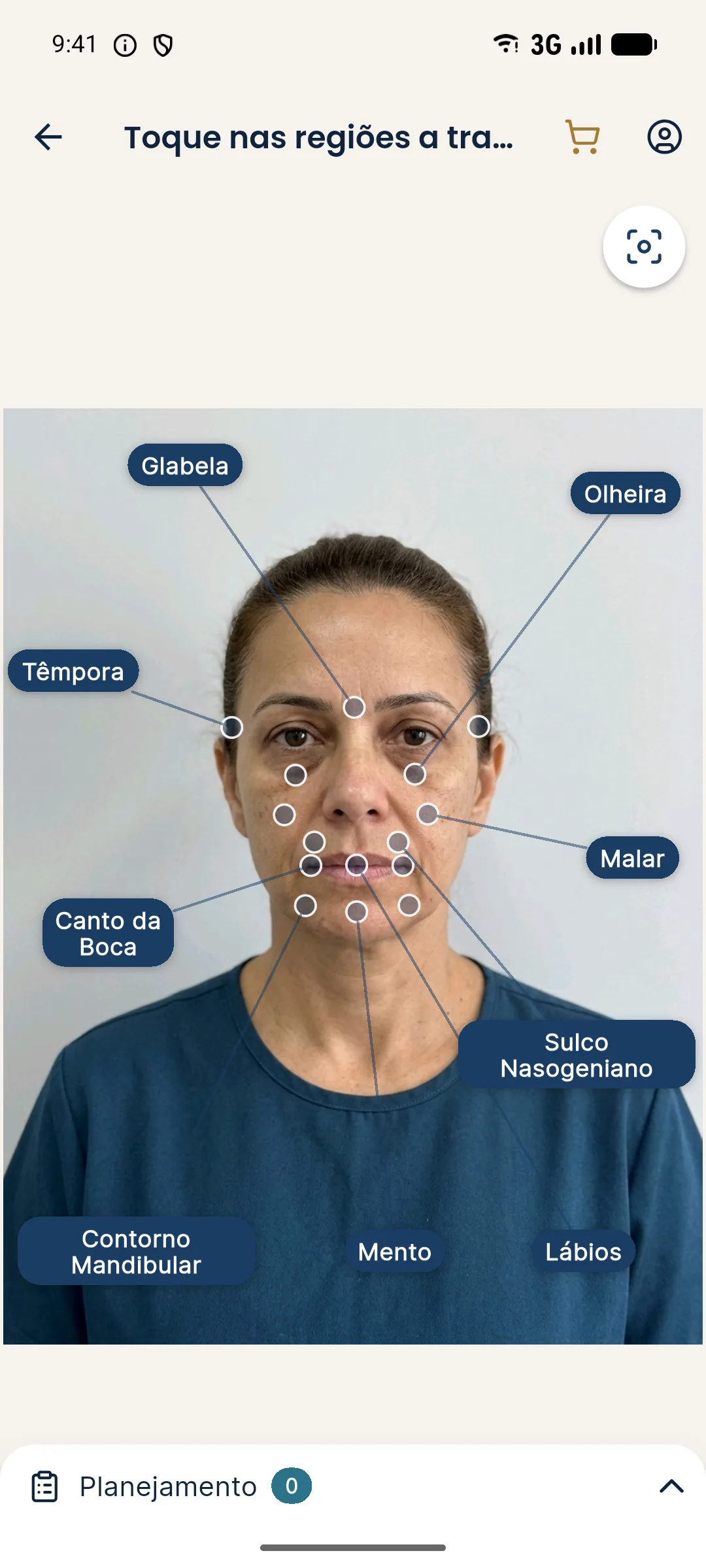
- Marking the areas. Where the plan will act, with the planned ml for each area.
- Simulation. An "after" image that illustrates the direction of the plan.
- Plan document. A written summary, quote and informed consent form.
What are the steps in practice?
The order matters. Whoever simulates before assessing risks letting the image drive the diagnosis, when it should be the other way around. The assessment comes first; the image is there to communicate.
What does it solve?
| Problem in the practice | How visual planning helps |
|---|---|
| The patient does not understand what will be done | They see the marked areas and the planned volume |
| Expectations above what is realistic | A simulation at a realistic intensity aligns what is possible |
| Forgetting the starting point | The standardized photo becomes a baseline |
| A quote that is hard to explain | Each area shows its ml and price |
| Changes of mind between sessions | The written plan and the image serve as a record |
This ties directly into how to use the frontal photo in the conversation and simulation and quote in the same conversation.
What stays outside visual planning?
This is the point that most needs to be clear. Digital planning does not:
- make a diagnosis (that is a clinical act of the professional);
- define technique, depth, injection point or product;
- measure with millimeter precision the result the face will have;
- predict swelling, individual response, hidden asymmetries or expression dynamics;
- replace informed consent or the form that documents it.
How does the frontal photo fit in, and what does it not show?
The frontal photo lets you assess symmetry, the proportion of the thirds, face width and midface volume. It does not show projection, the profile of the nose and chin, or the relationship between lip and chin from the side. So if your plan depends on the profile, the clinical assessment and the profile photo are still needed, even if the simulation uses only the frontal one. The step-by-step assessment is in facial analysis in facial harmonization.
What legal and ethical precautions come with it?
A photo of the face is sensitive personal data. Before photographing and before sending the image to any service, ask for specific consent and record it. In Harmy, the photo is sent to a third-party AI to generate the image, with the professional's acceptance in the app; the face contours are calculated on the device and are not stored. The responsibility for obtaining the patient's authorization lies with the clinic.
More details on consent and privacy in consent and LGPD in photos (LGPD is Brazil's data protection law) and in the ethics of simulation.
When is it worth adopting?
It is worth it when you notice the patient leaves the consultation without having understood the plan, when the quote is questioned for lack of clarity, or when expectations diverge from what you planned. It is not worth it as a substitute for a good assessment, or as a way to pressure the decision.
Start small: standardize the photo, record the concern, note the ml per area. Only then add the simulation. The goal is a clear plan for both sides, and technology is only useful if it makes the plan clearer.
How do you adopt it without overloading the routine?
The mistake of those who start is trying to roll everything out at once. Adopting in layers works better: first the standardized photo, then recording the concern, then marking and, last, the simulation. Each layer needs to be stable before the next one comes in, because each depends on the previous one to make sense.
Also define who does what. The photo can be taken by the team, as long as it follows the standard; the assessment and the decision on the plan always belong to the professional. A short script posted in the room (distance, height, light, neutral expression) reduces variation between visits and between team members.
How do you know it is paying off?
Without inventing numbers, look for qualitative signs: the patient repeats the question "but how will it look?" less, the quote is discussed with less friction, follow-ups become photo comparisons rather than memory, and the team knows where each record is. If none of this improves, review the workflow before blaming the tool.
What mistakes should you avoid in the first weeks?
- using the simulation before assessing the patient;
- leaving the image on screen while you are still explaining risks;
- photographing with different light and distance each time;
- storing photos on team members' personal devices;
- treating the digital plan as a substitute for the patient record.
Each of these mistakes has a simple, cheap fix. The most costly is the last one: the patient record remains the document of the visit, and digital planning is a visual complement to it.
Frequently asked questions
What is digital visual planning in facial harmonization?
It is the use of photography and digital tools, such as marking areas and simulation, to record the assessment, communicate the plan to the patient and align expectations before any injection.
Does digital planning replace the clinical assessment?
No. A photo is a two-dimensional snapshot. Palpation, dynamic assessment, medical history and examination remain the basis of the plan.
Do I need photos from several angles to plan?
For the clinical assessment, yes: frontal, profile and oblique views help a lot. Harmy's simulation uses only the frontal photo.
Can the digital simulation be shown as a guaranteed result?
No. It is an illustration of one possible direction. The actual result varies with the patient's response and with technique.
Harmy images are illustrative simulations generated by AI from the patient's photo to support the conversation between practitioner and patient. They are not a promise of results. Indication, technique, product and volume are solely the decision of the licensed practitioner. This content is for information only and does not replace a clinical assessment.