
Hollow Temples: Why It Happens and How to Treat It

 BeforeSimulation
- Highlighted area
Quick facts
- Question
- Why do the temples become hollow and how to treat them
- Usual causes
- Age, weight loss, temporal fat loss, genetics and muscle atrophy
- Treatment
- Volume replacement, in stages and at the professional's discretion
- Attention
- High vascular risk area, with superficial vessels and nerve branches
Anyone who has looked in the mirror and found the side of the forehead "hollow" knows the complaint. A hollow temple changes the frame of the face, makes the bone more prominent and creates a shadow under the hairline. This page helps you understand why it happens, how to assess the degree of loss and how to approach treatment with caution, since the area has particularities that call for respect.
Why does the temple sink?
The temporal region is a natural depression, the temporal fossa, filled by the temporalis muscle and a temporal fat pad, under the skin and fascia. Over time, this whole unit loses volume. The fat atrophies, the muscle can thin (even in people with little chewing use) and the bone also remodels.
Causes and associated factors that often come up in the medical history:
- Age: hollowing progresses slowly and is common from middle age onward.
- Weight loss: rapid or large weight loss removes facial fat, and the temple is often one of the first places to show it.
- Genetics and facial type: thin faces, more prominent bones and a pronounced brow ridge tend to show hollowing earlier.
- Use of certain medications or conditions that alter fat, evaluated by the professional.
- Smoking and sun exposure, which speed up aging of the skin and tissue.
How to assess the degree of loss?
| What to observe | Mild loss | Moderate to marked loss |
|---|---|---|
| Side line of the forehead | Smooth curve with a slight shadow | Visible concavity, bone line evident |
| Zygomatic arch | Barely evident | More prominent by contrast |
| At rest, from the front | Discreet shadow | Significant shadow under the hairline |
| In three-quarter view | Harmonious transition | Step between forehead and face |
| On palpation | Soft, full tissue | Palpable depression over the bone |
Assess with the patient from the front, in three-quarter view and with soft side lighting. A standardized photo helps measure progress, as described in standardized clinical photography. Another clue is the relationship between the temple and the cheekbone: sometimes the lack of continuity between the two areas is what bothers the patient.
What are the treatment options?
The general principle is to replace the lost volume, gradually. The choice of resource, volume and technique belongs to the licensed professional, in line with the rules of their professional board, and depends on the depth of the loss, skin thickness and what the patient wants.
- Hyaluronic acid filler: immediate effect, reversible with hyaluronidase, widely used in temple filler.
- Collagen biostimulator: gradual effect through tissue stimulation, described in biostimulator in the temples.
- Combined plan: in some cases the volume is replaced first and the transitions refined afterward.
- Autologous fat: a surgical option, indicated for selected cases and outside the scope of the orofacial harmonization (HOF) practice in many services.
The plan is usually staged, because the temple responds subtly and excess volume creates a "puffy" look that gives the treatment away. Volume ranges vary a lot and depend on the face.
Why does the area demand so much care?
The temple has superficial and deep vessels, such as the superficial temporal artery and branches of the middle temporal artery, as well as nerve branches that run through thin layers. There are communications with the orbit's territory, which explains why the area is among the high vascular risk zones. Details are in temple filler: risks and temporal anatomy for injectables.
Other possible effects: swelling that gathers by gravity and descends to the periorbital region, bruising, pain when chewing in the first days, asymmetry and irregularity. Warning the patient about swelling avoids panic on the second day.
What to tell the patient?
How Harmy helps with planning
In Harmy, mark the Temple area, set the volume in ml and the intensity (Subtle, Moderate or Pronounced) and generate the projection over the frontal photo. In a bilateral area, the ml is the total, divided between both sides. The projection is usually discreet, which fits the conservative planning the area calls for.
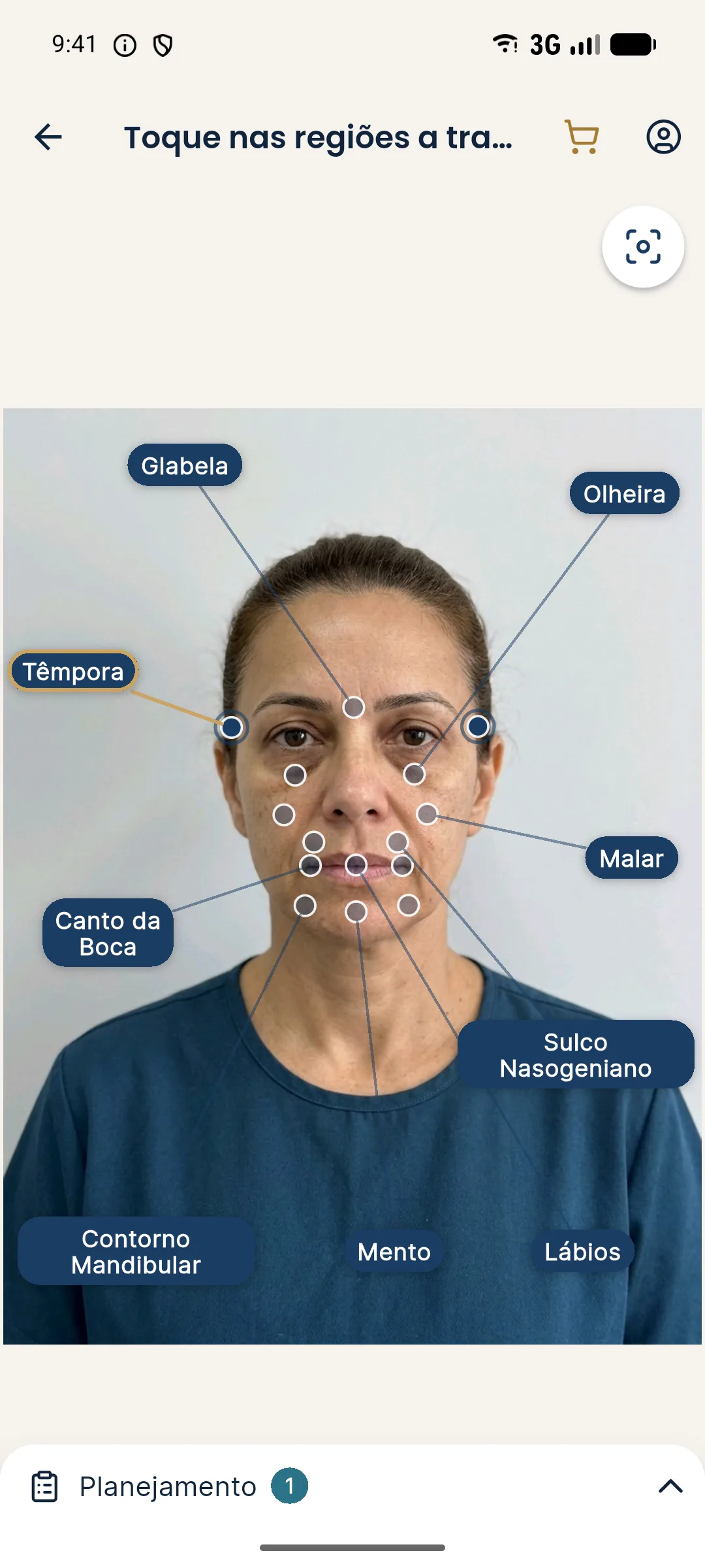
This is an illustrative simulation, not a promise of results, and the frontal photo shows lateral depth poorly. Use the slider to talk about contour, and assess the actual result from other angles with the patient present.
In short: a hollow temple is volume loss that builds up over time, and treatment is prudent replacement, in stages, by someone who knows the area's vascular anatomy.
Frequently asked questions
Why do the temples become hollow over time?
Because the temporal fat pad and the muscle lose volume, and the bone in the area also remodels with age. Weight loss, especially rapid, can accentuate the hollowing.
Do hollow temples make the face look older?
Hollowing often gives the face a more tired, drawn look, because the transition between the forehead and the face loses its smooth curve. That is a perception, and the plan should respect the patient's wishes.
How is a hollow temple treated?
Generally with volume replacement, using hyaluronic acid or a biostimulator, at the licensed professional's discretion. The plan is usually conservative and staged.
Is temple filler dangerous?
It is considered high vascular risk, because the area has superficial and deep arteries and veins. It requires anatomical knowledge, prudent volume and hyaluronidase on hand.
Harmy images are illustrative simulations generated by AI from the patient's photo to support the conversation between practitioner and patient. They are not a promise of results. Indication, technique, product and volume are solely the decision of the licensed practitioner. This content is for information only and does not replace a clinical assessment.