
Downturned outer eye corner: assessment and options

 BeforeSimulation
- Nerve
- Highlighted area
Quick facts
- Concern
- Lateral corner of the eye turned downward, sad or tired look
- Measurement
- Canthal angle, between the medial and lateral corners, on a standardized frontal photo
- Contributors
- Bone anatomy, cheekbone, lateral skin, eyebrow and muscle
- Limit
- Canthal surgery cannot be replaced with injectables
The lateral corner of the eye is one of the main contributors to expression: a higher corner conveys liveliness, while a downturned corner can give an impression of sadness or fatigue. When a patient complains of a "droopy eye" or a "sad look," the practitioner needs to know how much of that tilt is structural (and therefore fixed) and how much is soft tissue (and therefore modifiable).
This page shows how to measure, which layers contribute and what harmonization offers. It complements sad look, asymmetric eyes, gaze tilt and lateral canthus and tired look: assessment.
What is canthal tilt?
It is the angle between the line joining the medial and lateral corners of the eye and the horizontal. In many faces, the lateral corner sits slightly above the medial one, which contributes to a more "open" look. The canthal tilt varies considerably with sex, ethnicity, age and genetics, and there is no single number to use as a target.
What matters is the change relative to the patient's own pattern: a corner that has dropped over time, or that has always turned downward and now bothers them more. Compare with older photos, when available.
Which layers contribute?
| Layer | Role | Does facial harmonization change it? |
|---|---|---|
| Bone structure | Shape of the orbital rim and position of the canthal tendon | No |
| Cheekbone support | Projection of the midface below the eye | Yes, in part |
| Lateral skin and muscle | Orbicularis oculi, skin at the corner, crow's feet | Partially |
| Eyebrow | Position of the tail, which pulls on the look | Yes, in part |
| Upper eyelid | Ptosis or excess skin | Generally surgery |
| Temple | Volume loss that "pulls" the upper third | Yes |
The orbicular retaining ligament and the lateral canthal tendon are anchoring points that, when they loosen, allow the corner to drop. The structural part and the canthal tendon belong to the surgical field. For the eyelid, see drooping eyelid and eyebrow drooping at the tail.
How to assess
The exam needs a standardized photo, because tilt is very sensitive to head posture.
Pay attention also to the eyelid exam: an upper eyelid with excess skin or ptosis changes how the eye reads, and patients often attribute to the "corner" something that belongs to the eyelid. See sunken upper eyelid.
What harmonization can offer
The logic is to work on the soft tissue layers that contribute to the droop, at the discretion of the qualified practitioner.
Cheekbone support. Malar filler supports the midface and can reduce the impression of lateral droop of the eyes. In cases of tear trough and low cheekbone, this is the most discussed path. See sunken eyes and low cheekbone.
Temple. Temporal volume loss can contribute to droop of the lateral eye. Temple filler is an option, in an area of high vascular risk. See hollow temples and temples and eyebrow: combining in the plan.
Eyebrow. Toxin at selected points can change the position of the eyebrow tail and how the eyes read. See toxin for the eyebrow.
Lateral skin. The lines at the corner ("crow's feet") and skin quality respond to toxin and other resources. See pronounced crow's feet.
The limit: a corner that droops from canthal tendon laxity or significant excess skin is surgical. Facial harmonization does not replace canthopexy or blepharoplasty, and the patient should hear this clearly.
Risks
In the periorbital and temporal region, risks include swelling, bruising, asymmetry, nodules and, less often, serious vascular complications, with risk to vision. The area has branches of the superficial temporal and ophthalmic arteries, and hyaluronidase should be on hand. Toxin in the periocular region can cause ptosis, temporary diplopia and asymmetry. The consent form should name these risks.
How to show it in Harmy
In Harmy, the Cheekbone and Temple areas show the effect of support on the frontal photo, and the Tear Trough area shows the shadow under the eye. The projection changes only what was marked and does not alter the tilt of the eye corner: the app does not move structures, it only adds filler volume (toxin only in the glabella and the chin). This should be told to the patient.
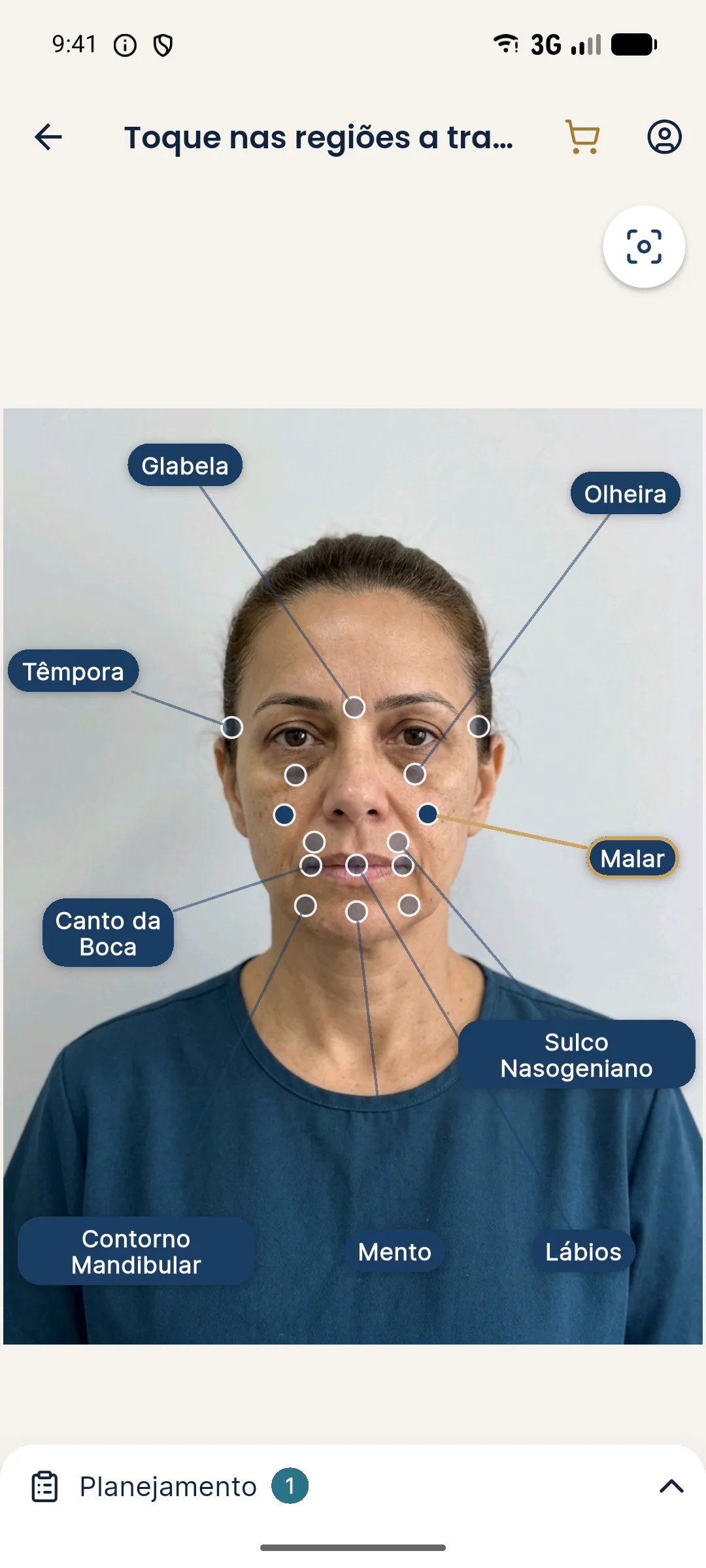
An honest approach is to make two projections (Cheekbone only, and Cheekbone with Temple) and compare. The patient sees how much support changes the whole and understands that the corner itself stays the same. The input photo should have the head straight and the gaze at the camera.
The look in the photo and in real life
Canthal tilt is greatly affected by photo details: camera height, chin elevation, light coming from above or below and even the type of lens. A photo taken from below makes the corner look lower, and a photo from above raises it. A patient who shows a social media photo to prove the concern may be seeing the effect of the angle.
So always do the assessment on the standardized photo and with the patient facing you in the office. Compare with their image in the mirror and on a video call. If the concern exists in all conditions, it has an anatomical basis. If it shows up in only a few, the conversation is about perception. Record this comparison and show it to the patient. This care avoids treating a concern that depends on the camera angle and reduces the risk of dissatisfaction later.
Wrap-up
Record the standardized photos, the angle measurement, the contributing layers and the limit explained. For the patient with eyes at different heights, see eyes at different heights and tilted bipupillary plane. Volumes and techniques are at the discretion of the qualified practitioner, according to the rules of their professional board.
Frequently asked questions
What is canthal tilt?
It is the angle formed by the line joining the medial and lateral corners of the eye and a horizontal line. In many faces considered youthful, the lateral corner sits slightly higher, but it varies with sex, ethnicity and the individual.
Is a downturned eye corner a facial harmonization concern?
In part. The droop can come from bone anatomy, cheekbone support, lateral skin, the eyebrow or the muscle. Harmonization works on some of these layers and has a limit on others.
Does filling the cheekbone lift the eye corner?
Supporting the midface can change how the eyes read and reduce the impression of lateral droop. The effect is indirect and limited, and depends on the cause.
When is surgery the answer?
When the droop is structural, with canthal tendon changes or significant excess skin. The decision involves specialist assessment and a frank conversation with the patient.
Harmy images are illustrative simulations generated by AI from the patient's photo to support the conversation between practitioner and patient. They are not a promise of results. Indication, technique, product and volume are solely the decision of the licensed practitioner. This content is for information only and does not replace a clinical assessment.