
Swelling Gets in the Way of Comparing Results: What to Do

Quick facts
- Topic
- Swelling and reading the result
- Effect
- Apparent volume larger than the real one
- Timing
- Set by the practitioner, depending on the procedure
- Caution
- Do not decide on a touch-up or dissolving while swollen
Two days after the procedure, the patient sends a photo: "it looks way too big." A week later: "the swelling is gone, but it looks like it disappeared." The same change is read in opposite ways because swelling alters the apparent volume, the definition of the contour and even the patient's mood. For anyone who plans and compares results, ignoring swelling is the fastest way to draw the wrong conclusions.
How does swelling change the reading?
Swelling (edema) is fluid accumulating in the tissue. After a procedure, it can increase apparent volume, blur the contour and create temporary asymmetry. With hyaluronic acid, the property of attracting water (hydrophilicity) also contributes to volume variation early on. Exact behavior depends on the product, the area, the technique and the patient, which is why the timeframe is not uniform.
Most common effects on the reading:
- Overestimating the volume, which creates fear of overdoing it.
- Losing definition, which erases the difference between areas.
- Creating asymmetry that resolves on its own.
- Making measurement harder, since contours change from day to day.
For the overview of expected effects and complications, see post-procedure instructions and edema.
When should you take photos?
The timeframe is the practitioner's decision, depending on the procedure and the area, and should be told to the patient before the procedure. The golden rule: never compare a projection, a plan or a touch-up against the swollen photo. The discussion on photo timing is in when to photograph the result after the procedure.
How do you avoid wrong decisions?
- State the timeframe on procedure day. Hand over the instructions in writing.
- Provide a channel for questions. A message can save an unnecessary visit. See 24-hour message after the procedure.
- Avoid assessing a touch-up before the timeframe. The temptation to "top up" is common.
- Record the immediate photo as swelling. Flag it in the patient record, so it is not mistaken for the result.
- Re-examine in person. The photo helps, but the exam decides.
How does swelling affect comparison with the simulation?
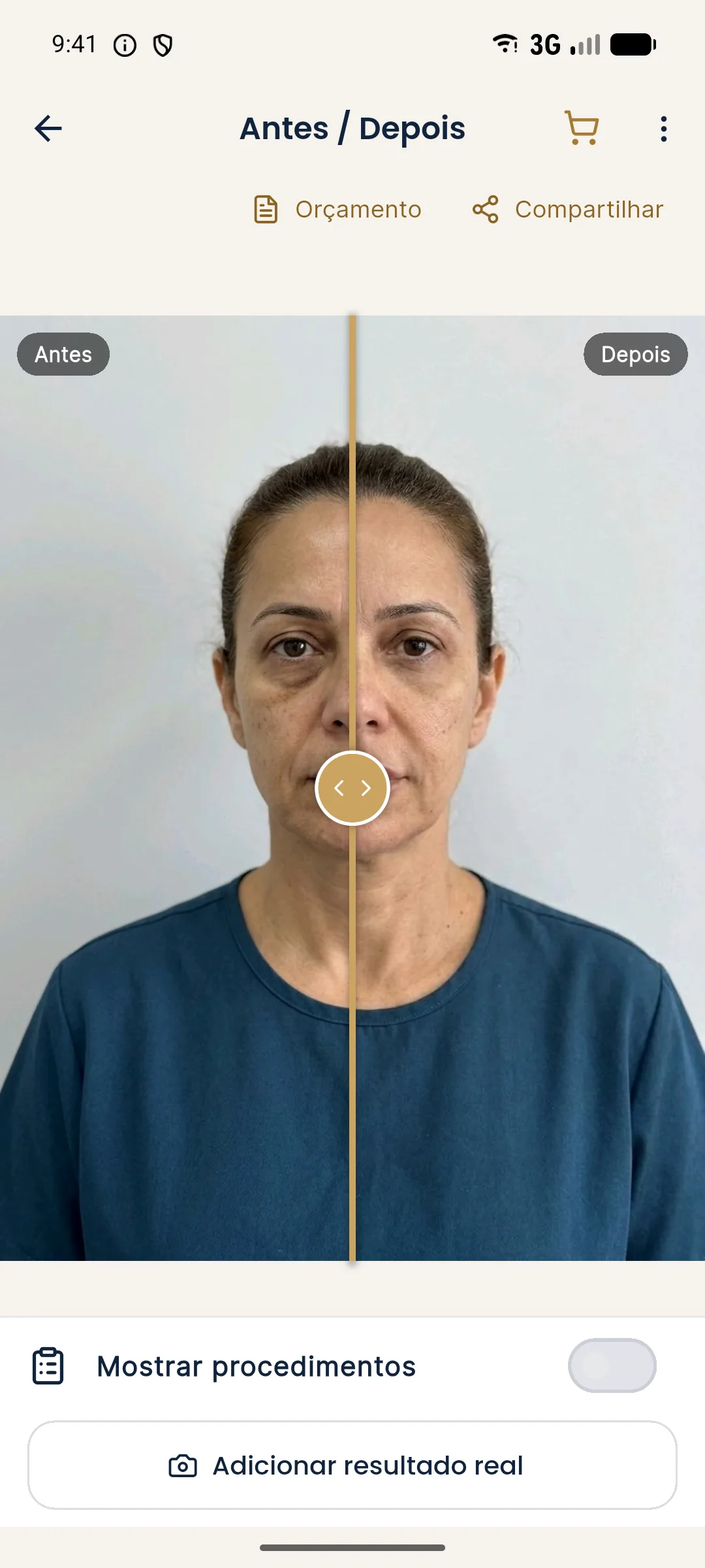
Harmy's projection is an illustrative image of a subtle change, without swelling. Comparing the projection with a photo of the swollen patient produces a difference that does not exist in the final result. That is why the "Projection vs Actual" comparison should be made after the swelling has resolved, with an aligned photo. See comparing projection and actual result and why the actual result differs from the simulation.
How do you explain it to the patient?
An anxious patient tends to check the mirror several times a day. The explanation should be concrete and short. Two ideas always hold: what they see is a result in progress, and the decision on a touch-up comes only later.
Communication about expected swelling is covered in explaining what expected swelling is and setting expectations for swelling and bruising.
What changes from area to area?
Swelling does not behave the same across the whole face. Loose-tissue areas, like the lower eyelid and the periorbital region, tend to accumulate fluid more easily and can show visible swelling even with a small volume. Lips usually swell markedly and quickly, and the patient is alarmed by the temporary asymmetry. Firmer-tissue areas, like the chin and jaw, tend to show less variation, but the contour may still be altered in the first days.
That is why the reassessment timeframe and the follow-up message should be area-specific. A plan with lips and the under-eye area, for example, may have two photo dates, each with its own logic. Telling the patient "we'll see in two weeks" without considering the area is an oversimplification that can create anxiety.
Some individual factors also weigh in: use of anticoagulants or anti-inflammatories, sleep, salt, alcohol, intense exercise right after the procedure and a personal tendency to swell. Post-procedure instructions cover these points and are in post-procedure instructions and gym after facial harmonization.
Record in the patient record the area, the swelling seen in the immediate photo and the date of the follow-up photo. This lets you better predict the patient's behavior in future sessions and guide them more precisely.
How do you support the patient during the swelling phase?
A short message the next day, asking how they are and reminding them of the photo date, reduces anxiety a lot. Reinforce the warning signs and the contact channel. Avoid generic automated replies: the patient wants to feel that someone is following the case.
Summary
Swelling is a confounding variable. Agree on the timeframe beforehand, record the immediate photo as such, do not decide on a touch-up while swollen and compare with the projection only after resolution. Warning signs call for immediate assessment. Management belongs to the qualified professional, in line with the rules of their professional board in Brazil.
Frequently asked questions
Does swelling get in the way of assessing the result?
Yes. While there is swelling, the apparent volume is larger and the contour less defined. The final assessment should wait until it resolves, within the timeframe the practitioner advises.
How long does swelling last after filler?
It varies with the area, the product, the technique and the patient. It is usually most evident in the first days and fades over days to weeks, at the discretion of the practitioner who schedules the follow-up.
Can I decide on a touch-up while the patient is still swollen?
Generally not recommended. Deciding under swelling can lead to unnecessary volume, so the approach is to wait and re-examine at the agreed time.
How do I explain swelling to the patient?
Tell them that part of what they see is temporary swelling, that the result settles over time, and agree on the date of the follow-up photo.
Harmy images are illustrative simulations generated by AI from the patient's photo to support the conversation between practitioner and patient. They are not a promise of results. Indication, technique, product and volume are solely the decision of the licensed practitioner. This content is for information only and does not replace a clinical assessment.