
Why the actual result differs from the simulation

Quick facts
- Core idea
- The simulation shows an intention in a still image; the actual result is a biological process that unfolds over time
- Patient factors
- Skin thickness and quality, fat, bone, muscle, prior asymmetry, hydration and habits
- Procedure factors
- Product, volume, site, technique and the timing of the assessment after the injection
- Safety phrase
- The image is an illustration, the result is individual and is assessed once the swelling has gone down
Every professional has heard the sentence: "it came out different from the photo". Sometimes the patient is right, sometimes not, and in both situations the explanation is the same: a simulation and a clinical result do not belong to the same category. One is a still image produced from a photo and some numbers. The other is a biological phenomenon, happening in living tissue, over days and weeks, with variables that no image captures.
This page organizes those variables into two groups, the patient's and the procedure's, so you have a clear vocabulary when explaining the difference before and after. The ethical basis of the presentation is in ethics of result simulation.
Do the simulation and the result measure the same thing?
No. The simulation answers the question "how would the face look if the chosen volume and intensity were applied to this contour?". The actual result answers "what happened in this patient's tissue after the procedure was done?". The first is a visual estimate; the second is the response of an organism.
In Harmy, the projection uses a frontal photo and changes only the marked areas. It does not see skin thickness, the bone behind it, the muscle in motion or the inflammatory response. The technical limits are described in limits of 2D simulation.
Which patient factors weigh the most?
These are the ones you assess at the consultation and that the image cannot represent. The main ones:
- Skin: thickness, elasticity and tendency to swell determine how the final contour looks. The relationship is covered in skin thickness in planning.
- Bone structure: bone defines the support. A bone deficit is not corrected by volume, a point covered in when bone is the problem and not volume.
- Fat and compartments: fat distribution varies from patient to patient and changes the effect of the same volume.
- Prior asymmetry: the starting face is already asymmetric, and the result inherits that asymmetry. See simulation for a patient with asymmetry.
- Habits and health: sun, smoking, sleep, weight and hydration influence tissue quality. The topic appears in habits that change the plan.
Which procedure factors weigh the most?
Here are the decisions you make that the image also fails to capture:
- Product: each product behaves in its own way in terms of water retention, firmness and integration into the tissue. The same number of ml can behave differently.
- Volume actually used: the planned ml is not always the ml injected. The difference is covered in planned ml vs used ml.
- Clinical choice of site and technique: decisions made by the licensed professional at the time of the assessment and beyond the reach of the image.
- Interval between sessions: a result built in two or three stages does not look like one from a single session. See interval between sessions in the plan.
How does swelling interfere with the comparison?
Swelling is the most underestimated factor in the comparison. In the first days, the treated area may be fuller than planned, with a contour different from what is expected in the final version. There is also bruising, which changes the color and the reading of the photo. The reverse also happens: as the swelling goes down, the result may look smaller than the image suggested, and the patient gets frustrated.
The date of the comparison photo must be set beforehand. The window depends on the procedure and the patient, and the choice is up to the professional. The topic has its own page in when to photograph the result after the procedure and in swelling gets in the way of result comparison.
What can the difference in photo capture explain?
Half of the "difference between simulation and reality" is actually a difference between two photos. If the result photo was taken with different light, distance, expression or angle, the contour looks different even with no real change. The most common causes are in alignment mistakes when comparing photos and in distance and lens in the assessment photo.
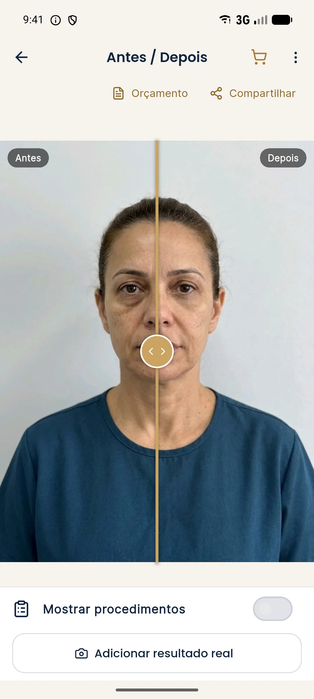
In Harmy, the Actual result stores the post-procedure photo aligned by the eyes. Guided capture shows the "before" photo in transparency over the camera, which helps repeat the framing. The app compares Projection, Actual result and Projection vs Actual.
How do you explain the difference to the patient at the consultation?
The best time is before the procedure. The preventive conversation takes two minutes and avoids a difficult conversation later. A supporting script:
These pages complement the script: explaining that a simulation is not a guarantee and simulation to calibrate expectations.
And what if the difference really comes from the procedure?
Not every difference is perception. If, once the swelling has passed and the photo is taken in the standard, the result departs from the plan, treat it as a clinical finding: reassess, record and talk. There may be new asymmetry, insufficient volume or unexpected distribution. The approach is up to the licensed professional and follows the council's rules. Paths for the follow-up are in what to reassess at the plan follow-up and in re-planning after the first session.
How do you turn this explanation into a practice routine?
Standardize three moments. At the consultation, say that the image is illustrative and note that you said it. After the procedure, book the comparison follow-up before the patient leaves. At the follow-up, photograph in the standard and compare side by side, with the patient. This routine reduces the feeling of surprise and shows that your judgment is not opinion, it is method.
Frequently asked questions
Why did my result turn out different from the simulation?
The simulation is a still image of an intention. The actual result depends on tissue, product, volume, technique, swelling and the day it is assessed, so it may be more discreet or differ in details.
Does swelling change how the result is assessed?
Yes. In the first days, swelling can increase or distort the contour, and the most honest assessment usually happens once it has gone down, on a date set by the professional.
Can the simulation be treated as a commitment to a result?
It should not be. It is an illustration to support the decision, and this must be said in the conversation and in the consent form, according to the rules of your professional council.
How do you explain the difference without discouraging the patient?
By showing the image as a direction, not a destination, and by arranging a follow-up where you compare photos in the same standard and decide together whether any adjustment is needed.
Harmy images are illustrative simulations generated by AI from the patient's photo to support the conversation between practitioner and patient. They are not a promise of results. Indication, technique, product and volume are solely the decision of the licensed practitioner. This content is for information only and does not replace a clinical assessment.