
How to Compare the Projection and the Actual Result in HOF

Quick facts
- Topic
- Comparison between projection and actual result
- In Harmy
- Projection, Actual result and Projection vs Actual
- Before comparing
- Wait for the swelling to subside and align the photo
- Caution
- The projection is illustrative, not a target
Comparing the projection with the actual result is one of the most delicate moments of follow-up. If the patient sees the two images and notices a difference, they tend to think something went wrong. If they see no difference, they tend to think the simulation "promised and delivered". Neither reading is the right one. The comparison serves to calibrate the conversation, not to give a grade. This page offers criteria for doing it methodically.
What can and cannot the comparison conclude?
The Harmy projection is an illustrative image, generated from a frontal photo and the volumes and intensities marked. The actual result is the outcome of a procedure done on a living face, with tissues that react. The two images are different in nature, and so the comparison:
- Can show whether the direction of the change (where and how much) resembles what was agreed.
- Can support a conversation about what the patient notices and what they still want.
- Cannot prove that the procedure was done well or badly.
- Cannot serve as a guarantee for the next plan.
The difference between the two is detailed in why the actual result differs from the simulation and in difference between simulation and actual result.
When should you photograph the actual result?
The right moment avoids comparing swelling. Guidance on timing depends on the procedure, the product and the area, and rests with the professional who performed the treatment. In general terms, swelling can change volume and contour in the first days, and the most faithful reading comes after it subsides. See when to photograph the result after the procedure and swelling gets in the way of comparing results.
Which criteria should you use?
The table below organizes the comparison along axes the professional controls.
| Criterion | What to check |
|---|---|
| Alignment | Face in the same position, distance and tilt |
| Light | Same direction, intensity and color |
| Expression | Neutral in both images |
| Timing | Photo taken after the swelling period |
| Area | Compare only the areas treated |
| Perception | What the patient sees and what the professional sees |
| Concern | Has the original concern improved, in the patient's view? |
The typical alignment errors are in alignment errors when comparing photos, and the capture technique in Harmy is in aligned follow-up photo in Harmy.
How does Harmy help with the comparison?
In Harmy, in the patient's session, you can use the "Add actual result" option. The app saves the follow-up photo aligned by the eyes, and guided capture shows the "before" photo in transparency over the camera so you can reproduce the pose. Afterward, the comparison offers three modes: Projection, Actual result and Projection vs Actual.
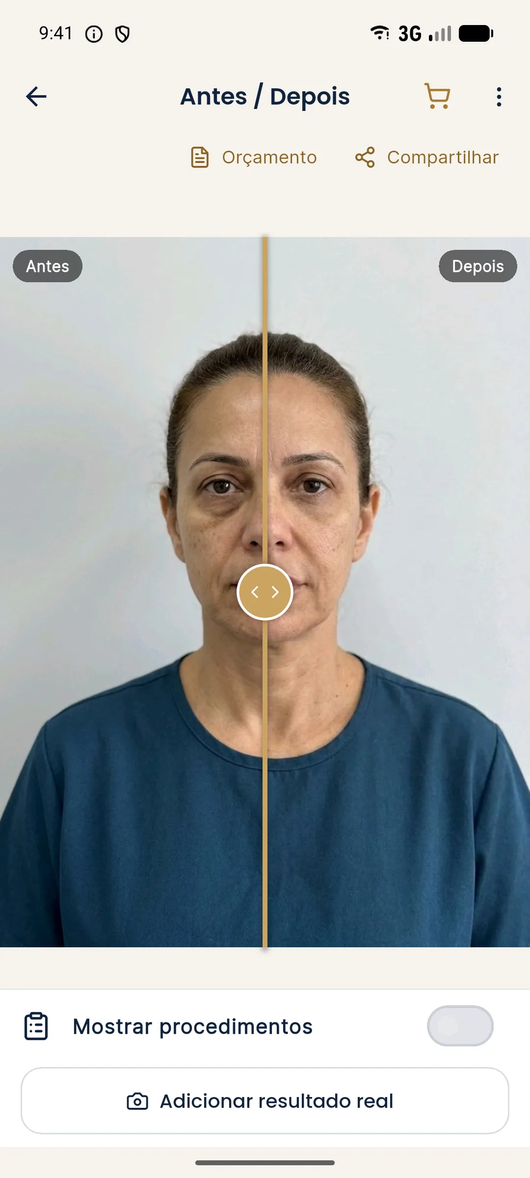
The "Projection vs Actual" comparison is for the professional to look at the direction of the changes together with the patient. It does not calculate accuracy or measure volume. Read how the feature works in comparing projection and actual result in Harmy and in history and actual result.
How do you lead the conversation?
The opening sets the tone. Start by asking, not explaining.
If the patient is satisfied, record it and schedule maintenance. If they are dissatisfied, listen to the end, confirm the concern and assess whether an adjustment is indicated. If the patient is satisfied but you see a problem, discuss it carefully: it may be a chance to guide them on maintenance or limits. The structure for responding to a result below expectations is in communicating a result below expectations.
And when the difference is large?
Consider before concluding:
- Is the photo aligned and within the time frame? If not, retake it.
- Was there swelling, a bruise or a complication? See replanning after a complication.
- Was the ml performed what was planned? Record the deviation, as in planned ml vs. ml used.
- Was the expectation calibrated at the consultation? See simulation to calibrate expectations.
- Has the concern changed? Sometimes the patient starts noticing another spot. See replanning after the first session.
What should you record after the comparison?
A comparison without a record gets lost. At the end of the conversation, note in the patient record:
- The date of the result photo and the time elapsed since the procedure.
- Whether the capture followed the standard (distance, light, expression, alignment).
- The patient's perception, in their own words, of what changed and what still bothers them.
- Your clinical assessment, kept separate from the patient's perception.
- The decision: maintain, wait, adjust or close the plan.
- Consent to store the images and for any use beyond the patient record.
This record helps calibrate the same patient's next plans. If the projection was consistently more evident than the actual result, use Subtle intensity in upcoming conversations. If the patient was satisfied with a result less evident than the image, that is a sign the expectation was well calibrated.
It is also worth observing what did not change: concerns the patient had reported that remain may call for a next stage, or a conversation about limits. Finally, remember that patient images are sensitive data. Use for advertising requires specific authorization, as in separate consent for image publication.
When should the comparison not be done?
There are times when it is better to postpone. If the patient has obvious swelling, if the result photo was taken under conditions very different from the before photo, or if there is suspicion of an ongoing complication, the comparison can be confusing. In those cases, deal with the clinical matter first and schedule a new capture.
Summary
Compare at the right time, with an aligned photo and equivalent light. Treat the projection as an illustration, not a target. Record what the patient perceives, adjust if there is a concern and make no promises. Every decision to adjust belongs to the licensed professional, after assessment, according to the rules of their professional board in Brazil.
Frequently asked questions
Should the projection match the actual result?
No. The projection is an AI-generated illustration based on the frontal photo and the volumes marked. The actual result depends on the product, the technique, the anatomy and the individual response.
When should I photograph the actual result for comparison?
After the period in which swelling usually subsides, within a time frame set by the professional according to the procedure. Photographing too early compares swelling, not results.
How does Harmy compare the projection and the actual result?
In the patient's session, the option to add the actual result saves a follow-up photo aligned by the eyes, and the comparison shows Projection, Actual result and Projection vs Actual.
What should I do if the actual result turned out different from the projection?
Record it, explain that the projection is illustrative and assess with the patient whether there is a concern. If there is, discuss the possible adjustments, without promising anything.
Harmy images are illustrative simulations generated by AI from the patient's photo to support the conversation between practitioner and patient. They are not a promise of results. Indication, technique, product and volume are solely the decision of the licensed practitioner. This content is for information only and does not replace a clinical assessment.