
Smile analysis in HOF: what to assess before planning
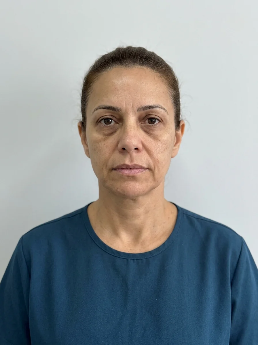
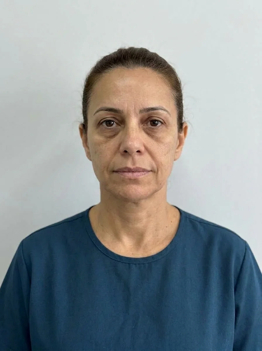 BeforeSimulation
- Nerve
- Highlighted area
Quick facts
- Goal
- Assess the smile at rest, in a soft smile and in a broad smile before deciding on lips, mouth corners and toxin
- Items
- Tooth display, smile arc, buccal corridor, symmetry, mouth corners and upper lip
- Why it matters
- Filling or paralyzing without looking at the smile can alter what the patient uses most every day
- Record
- Photo at rest, soft smile and broad smile, plus a short speech video
The smile is the first expression a patient uses to introduce themselves, and the one they most fear changing. Filling the lips, working on the mouth corners or injecting toxin in the lower third without understanding how that smile works means accepting the risk of altering something they do hundreds of times a day. Smile analysis is therefore part of the assessment, not an optional item.
This page lists what to assess, at rest and in movement, what to record and how the finding changes the plan. It fits into the sequence of facial analysis in HOF: step by step and dynamic facial analysis.
Why assess the smile at three moments?
A face has at least three clinical smiles, and each shows something different.
| Moment | What it shows | What to record |
|---|---|---|
| Rest | Display of the upper incisors with the lip relaxed, position of the lips and mouth corners | Frontal and profile photo, mouth relaxed |
| Soft smile | Social smile, the most frequent | Frontal photo with a natural smile |
| Broad smile | Maximum display, buccal corridor, gums, folds and mouth corners | Frontal photo with a spontaneous smile |
Incisor display at rest is one of the most useful indicators. It varies with age, upper lip length and tooth position, and tends to decrease with aging, as the upper lip lengthens and the lower teeth show more. What this means for the plan is in the lips view.
What should you look at in the smile, item by item?
The smile arc and the buccal corridor have their own pages, because the reading of each is detailed. The point here is that all of them go into the photo and the record, before any decision.
How does the smile change the lip plan?
The lip is the most visible element of the smile. Changing its volume can change tooth display and the harmony with the chin. A few examples of how the finding changes the reasoning:
- Short upper lip, with little tooth display. Volume in the upper lip can reduce display even further. The plan tends to be more conservative and prioritize balance.
- Long, thin upper lip. Assessing the ratio between the upper and lower lips and examining the Cupid's bow help decide where the volume fits.
- Smile with a lot of gingival display. The plan may involve assessing the upper lip elevators, in a context other than filler, as the professional sees fit.
- Overprojected lower lip. More volume there can accentuate the imbalance with the chin.
Lip proportion is a reference, not a target. The relationship between the upper and lower lip varies between people and between populations, and the ideal on paper is almost never the patient's ideal. The reading of proportion is in lips and chin: combining them in the plan.
What about the mouth corners and the smile muscles?
The mouth corners descend over time, and the modiolus, the meeting point of several muscles beside the corner, plays a role in this dynamic. The zygomaticus major pulls the corner up and outward when smiling. The orbicularis oris closes and projects the lips, and forms the vertical perioral lines.
In the assessment, observe:
- whether one corner rises more than the other when smiling;
- whether there are marionette lines (the line that runs down from the corner) at rest;
- whether the smile has always been asymmetric or only recently;
- whether there are lines that deepen when smiling.
The topic of mouth corners and marionette lines appears in lips and mouth corners: combining them in the plan and in mouth corners, marionette lines and chin: combining them.
How do toxin and filler change the smile?
Describing technique is not the role of this page. But it is worth knowing what the clinical literature describes at review level: toxin in the upper lip or near the zygomaticus can alter the smile; filler in the fold and the mouth corners can change the position of the corner; volume in the chin changes the support of the lower lip. Planning needs to anticipate these cross effects, and the conversation with the patient should include them. See also botulinum toxin for gummy smile.
What are the most common recording mistakes?
- Photo only at rest. It misses the dynamics and underestimates the effect on the mouth corners.
- Forced smile. A posed smile differs from a spontaneous one; ask for the spontaneous one.
- Photo without leveling. A tilted head simulates a buccal corridor asymmetry.
- Ignoring the dental midline. A dental midline deviation can look like a lip deviation.
- Not photographing the before. Without a record, any later complaint becomes an argument.
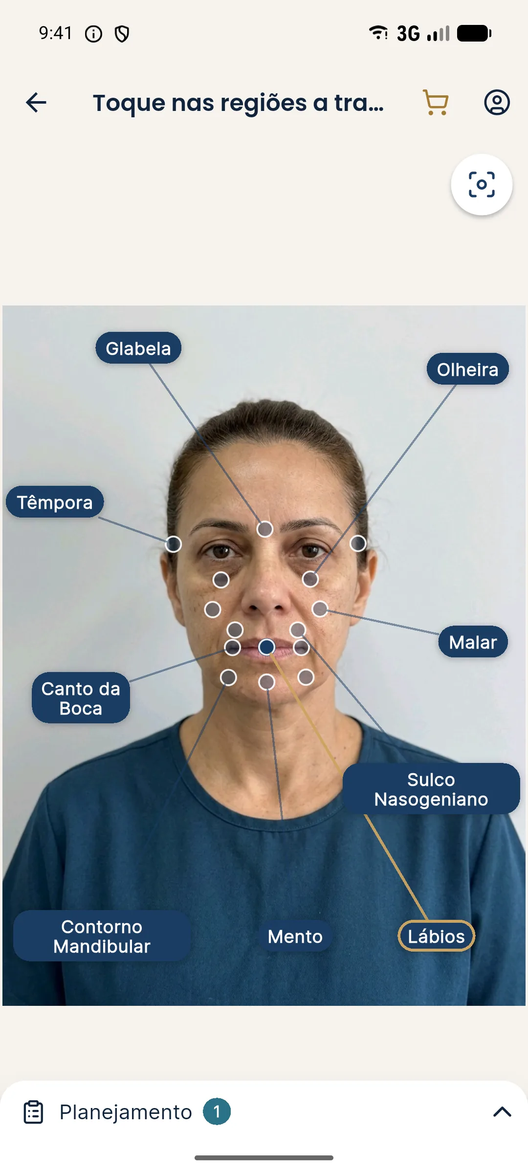
The app uses the frontal photo you upload. Photograph the patient with the expression you intend to analyze.
What should you tell the patient?
Myth or fact?
Summary
- Assess the smile at rest, soft and broad, and record all three.
- Tooth display, arc, buccal corridor, mouth corners and upper lip change the plan.
- Lip volume and toxin in the lower third have a cross effect on the smile.
- Document preexisting asymmetry before any procedure.
- The app's projection does not change the expression in the photo.
Frequently asked questions
What should I assess in the smile before filling the lips?
Tooth display at rest and when smiling, the smile arc, the buccal corridor, the symmetry of the mouth corners and the length of the upper lip. This defines the volume and where it fits.
Why photograph the smile in addition to the resting face?
Because the lip and the mouth corner change position when the patient smiles. What looks balanced at rest can become asymmetric or heavy in the smile.
Does a gummy smile affect planning?
It does, because a short upper lip or very active elevators change the approach to the lips and toxin. The assessment must be done in movement and the decision belongs to the licensed professional.
Does Harmy's projection show the smile?
The projection uses the frontal photo you upload and does not change the expression. To assess the smile, the planning photo needs to show the patient smiling or at rest, depending on what you want to analyze.
Harmy images are illustrative simulations generated by AI from the patient's photo to support the conversation between practitioner and patient. They are not a promise of results. Indication, technique, product and volume are solely the decision of the licensed practitioner. This content is for information only and does not replace a clinical assessment.