
Nasolabial fold different on one side: assessment
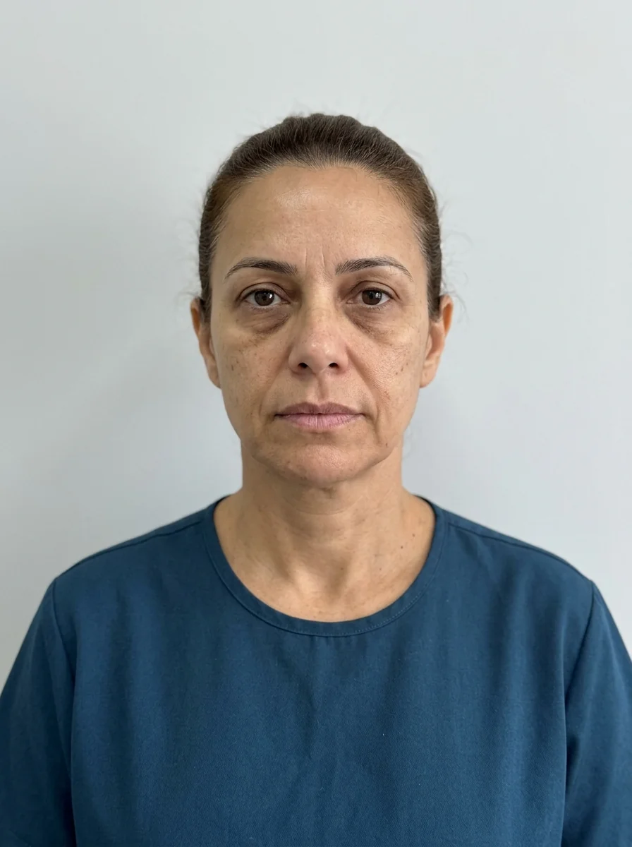
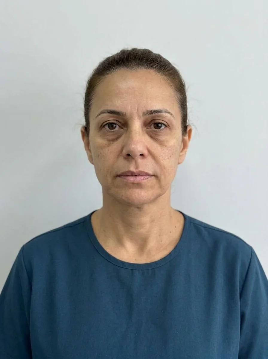 BeforeSimulation
- Highlighted area
Quick facts
- Concern
- Nasolabial fold visibly more marked, longer or more curved on one side
- Key question
- Does the difference come from soft tissue, skeleton, dentition, or the habit of chewing and sleeping on one side?
- Rule of thumb
- Treat the cause of the difference, not just even out the fold with different volumes
- Plan risk
- Correcting the deeper side with more product and producing a face that is more asymmetric in motion
The patient points to the mirror and says: "look, on the right side it's much deeper". They are right more often than you would think, because the human face is naturally asymmetric, and the nasolabial fold (smile lines) is one of the structures where asymmetry shows most. The question for the person doing the assessment is whether the difference deserves treatment, what produces it and whether treating one side differently from the other is the right decision.
This page separates the fold's asymmetry into layers, so the plan does not boil down to "put more product where it is deeper". The overview of the topic is in deep fold from nose to mouth and the fold from the procedure's point of view is in nasolabial fold filler.
Why the two sides are rarely equal
The nasolabial fold is the crease that separates the cheek from the upper lip, and its appearance depends on many things at once: the position and volume of the neighboring fat compartment, the bone of the maxilla, skin thickness, the action of the smile muscles and the position of the teeth. Each of these factors can differ from one side to the other.
That is why the same concern, a different fold, has a different origin in each patient. Good practice is to name the dominant cause before thinking about product. And to recognize that, even after treatment, the face will still have some asymmetry, which is normal and expected. On this, see explaining natural asymmetry to the patient.
The four layers of the difference
| Layer | How it shows | What to investigate |
|---|---|---|
| Bone | Maxilla or canine fossa more recessed on one side, greater shadow | Frontal and three-quarter view, palpation, dental assessment |
| Fat and support | Nasolabial compartment and cheekbone lower on one side | Compare the midfaces, cheekbone projection and ptosis |
| Muscle and dynamics | Smile higher on one side, more intense crease when smiling | Photo at rest and smiling, short video |
| Skin and habits | Marks from sleeping on one side, different tone, uneven sun exposure | Medical history of sleep, chewing, smoking, sun |
The fold at rest and the fold in the smile can tell different stories. At rest, the difference tends to reflect bone and fat. In the smile, it includes muscle dynamics. Compare both situations and record them.
When the cause is the skeleton
If the difference comes from one side of the maxilla being more recessed, the fold on that side looks deeper because the base is lower. Filling the fold compensates the appearance but does not change the structure, and the same amount of product on the other side would be too much. In certain cases, the decision is to project the midface on that side, leaving the fold for later, when the shadow has already decreased.
If the patient has a history of orthodontics, orthognathic surgery or crossbite, the conversation goes through that context. For a patient who complains of an uneven face overall, see asymmetric face and crossbite and asymmetry.
When the cause is habit
Chewing on one side, always sleeping on the same side and the habit of resting the face on a hand can contribute to differences in tone and skin marks over the years. It is a plausible factor, but hard to prove in an individual case, and so it should be presented to the patient as a hypothesis. See asymmetry from chewing on one side.
Guidance about habits can be given without promising that changing them will correct the fold. The expected gain is avoiding worsening and helping the treatment result last.
What to treat and what not to treat
The central decision is whether the goal is to even out the sides or to improve each side. Both are legitimate, but they are not the same thing. Evening out requires different doses per side and a frank conversation about the asymmetry that will remain. Improving each side respects the face as it is and tends to give a more natural result.
A prudent sequence is the following:
- Classify the dominant cause and note the layer involved.
- If cheekbone support differs, discuss the midface before the fold. The combined reading is in nasolabial fold and cheekbone in the plan.
- If the difference is dynamic, assess the movement before choosing the volume.
- If the fold is going to be treated, start with modest amounts and reassess each side separately.
- Schedule a follow-up and compare with standardized photos.
Safety in this area
The nasolabial fold is an area of vascular risk, because of the facial artery and its branches. Possible complications include swelling, bruising, nodules, asymmetry, irregularity and, rarely, vascular occlusion. Keep hyaluronidase available, know the warning signs and review vascular occlusion in the nasolabial fold. When the difference between the sides calls for different amounts, record the reason in the patient record.
Biostimulator and other options
In patients with loss of support on one side, a biostimulator in the midface or around the fold can be discussed, always at the professional's discretion and with an explanation of response time and the risk of nodules. Read biostimulator in the nasolabial fold and PDO threads in the nasolabial fold to learn the indications and limits.
How to use Harmy in this conversation
Harmy starts from a frontal photo and changes only the marked area. For a concern about an uneven fold, the app serves to show the patient what a volume on each side can do in the photo, but it does not create a symmetry the face does not have. This is useful in the conversation, because the patient sees that the difference between the sides is still there.
Because the ml in a bilateral area is split between the two sides, the app's projection does not replicate a different dose per side. This must be said to the patient, so the simulation does not turn into a promise of results.
Questions to test the reasoning
What to record in the patient record
Note the description of the asymmetry in the patient's own words, the standardized photos of each side, the dominant cause you considered, the amounts planned per side and the justification. Also record that the patient was informed that some asymmetry will remain and that the result depends on individual factors. For the photo standard, see standardized clinical photography. On common patient questions, natural facial asymmetry.
Frequent mistakes
The first mistake is evening out what cannot be evened out. Asymmetry is part of the face, and trying to zero it with product tends to produce excess volume on the deeper side and a mask-like look.
The second mistake is treating the fold without looking at the midface. When the base drops, the fold follows, and correcting only the fold ends up insufficient or exaggerated.
The third mistake is photographing without standardization. A slight head tilt or light coming from one side creates differences that do not exist. Before deciding, repeat the photo under controlled conditions.
Summary for the decision
A nasolabial fold that differs on one side is almost always natural asymmetry, with more than one layer involved. Classify the dominant cause, assess the midface, observe the movement, and only then decide whether, where and how much to treat on each side, with conservative amounts and a follow-up scheduled. Explain that some asymmetry will remain, document, keep hyaluronidase available and always act at the discretion of the qualified professional, in line with the rules of their professional council in Brazil, without promising results.
Frequently asked questions
Is it normal to have a nasolabial fold that differs from one side to the other?
Yes. Mild asymmetry is very common and can come from bone, teeth, muscle, skin or habits. The clinical question is whether the difference bothers the patient and whether there is a treatable cause, not whether it exists.
Does filling only the deeper side fix the difference?
Sometimes it improves the shadow, but it may not correct the cause, especially if the difference comes from cheekbone or maxilla support. The midface on each side is generally assessed as well.
Does sleeping on one side or chewing on one side change the fold?
Over the years it may contribute to differences in skin and tone between the sides. It is a factor to investigate in the medical history, not a guaranteed explanation.
Does the photo show the fold as more different than it is in real life?
It can. Head tilt, side light, smiling and the lens all change the appearance. That is why assessment uses standardized photos and in-person observation.
Harmy images are illustrative simulations generated by AI from the patient's photo to support the conversation between practitioner and patient. They are not a promise of results. Indication, technique, product and volume are solely the decision of the licensed practitioner. This content is for information only and does not replace a clinical assessment.