
Prioritizing by Anatomical Risk in a Facial Harmony Plan

- Highlighted area
Quick facts
- Principle
- Anatomical risk sets the safety schedule: how, when and with what preparation each area enters the plan
- Areas of concern
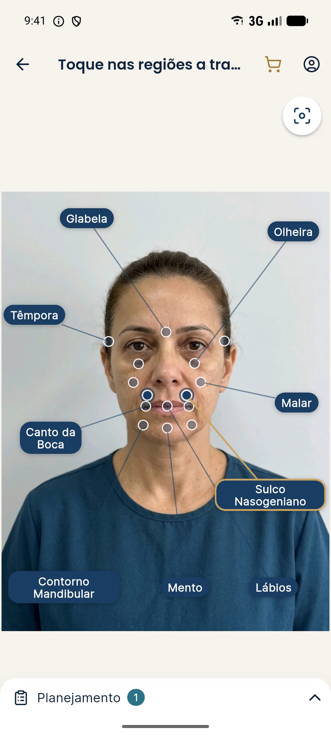
- Glabella, nose, nasolabial fold, temples and tear trough, because of vascular risk
- Mandatory
- Hyaluronidase on hand whenever hyaluronic acid is used, and a patient informed of the risks
- Caution
- This page covers planning and does not teach injection technique
No harmonization procedure is free of risk, and the areas of the face are not equal in this respect. Facial vascularization is rich and variable, with superficial branches and anastomoses between different systems. In some areas, a vascular complication can have serious consequences, which changes how the professional plans the order and the preparation.
The subject here is planning: how anatomical risk enters prioritization, scheduling and the conversation with the patient. There is no description of injection technique. The reader is a professional, but a public page is not a protocol.
Why does risk need its own place in prioritization?
Because visual impact and budget know nothing about anatomy. A plan ranked only by perceived improvement may put a high-risk area first, simply because it is the patient's concern, without the professional having thought about preparation, timing and specific consent. Prioritizing by risk works as a second filter, applied before the plan is presented.
Where the two logics meet is covered in prioritizing by visual impact in the plan. When they conflict, safety wins, and the patient needs to understand why.
Which areas are usually treated as areas of concern?
The safety literature on facial harmonization consistently points to a group of higher-caution areas. This is not an official classification, and assessment is individual.
| Area | Why it is an area of concern (at review level) |
|---|---|
| Glabella | Supratrochlear and supraorbital vessels, connected to the ophthalmic artery system |
| Nose | Dorsal and columellar arteries, with anastomoses and thin skin |
| Nasolabial fold | Close to the facial and angular arteries, which connect with the orbital territory |
| Temples | Superficial and deep temporal vessels, with anatomical variations |
| Under-eye area and tear trough | Very thin skin, nearby vessels and proximity to the eye |
To study the vessels themselves, there are dedicated pages, such as angular artery, supratrochlear artery and danger triangle of the face. They are an anatomy review and do not replace the professional's training.
How does risk enter the schedule?
There is no universal order, but there are principles that planning usually follows:
- Lower-risk structure first, when clinically appropriate. Results in the foundation often reduce the need to intervene in risk areas, because the detail improves with support.
- First approach to a risk area in a session with no other variables, so any complication can be interpreted clearly.
- Limited number of areas per day, a topic covered in how many areas to treat per day.
- Adequate interval between sessions in the same area, as in interval between sessions in a facial harmonization plan.
- Contingency plan defined beforehand, as described in contingency plan in facial harmonization.
What changes in consent for a risk area?
Specificity. A generic form saying "complications may occur" is not enough for areas where a complication is rare but serious. The patient has the right to know the name of the risk, the warning signs and what to do if they notice any. This is in line with good practice for informed consent and the duty to inform.
How to word the conversation is covered in communicating risk to the patient and in explaining areas of caution to the patient.
How does risk priority coexist with the patient's concern?
It is common for the main concern to be in an area of concern, such as the fold or the under-eye area. In that case, the plan can take one of three approaches:
- Treat first, with reinforced preparation, if the assessment shows a good indication and the professional has experience with the area.
- Treat the structural cause first, leaving the risk area for a second stage. This is the logic of treating the fold or the cause of the fold.
- Refer or postpone, when there is a contraindication, an unrealistic expectation or a limit of competence. The ethical reasoning is in when to say no to an aesthetic plan.
The decision among them is clinical, and the patient takes part with information, not pressure.
What changes when the patient has had procedures before?
Quite a lot. Previous surgeries, permanent products or products of unknown origin, and earlier complications change the expected anatomy and the risk. These details must be actively sought in the medical history, and the plan should allow for the possibility of not treating. Related topics include plan to correct prior excess and replanning after a complication.
How to balance safety and commercial communication?
Safety cannot become a sales argument, nor be left out to make the decision easier. Explain the risk as clearly as you explain the benefit. And avoid expressions such as "risk-free" or "no worries", which good practice and advertising rules discourage. What may be advertised, and how, is in what you may advertise in Brazil: CFO and CONAR. The overview of risks is in safety and complications: overview.
Frequently asked questions
Which areas of the face carry the highest vascular risk?
The glabella, nose, nasolabial fold, temples and tear trough are cited as areas of concern because of superficial vascularization and anastomoses that can lead to serious events.
Does prioritizing by risk mean treating the riskiest areas first?
Not necessarily. It means deciding more carefully when and how they enter the plan, and they often come after structural, lower-risk areas, at the professional's discretion.
Does the patient need to know the risks of each area?
Yes. Informing the patient of possible risks and complications is part of informed consent, in plain language and according to the rules of your professional board.
Does Harmy's simulation reduce risk?
The simulation does not change the risk of the procedure. It helps with planning and conversation, but safety depends on clinical assessment, anatomy and the conduct of the qualified professional.
Harmy images are illustrative simulations generated by AI from the patient's photo to support the conversation between practitioner and patient. They are not a promise of results. Indication, technique, product and volume are solely the decision of the licensed practitioner. This content is for information only and does not replace a clinical assessment.