
Prioritizing by visual impact in the facial harmonization plan

Quick facts
- Core idea
- Not every area changes the face in the same proportion; visual impact guides which comes first
- Three questions
- What does the patient notice? What does an observer notice? What supports the rest of the face?
- Rule
- Structure before detail: areas that support or project usually weigh more than refinement areas
- Caution
- Visual impact does not authorize skipping the safety order or inflating volume to draw attention
In a harmonization plan with several possible areas, the practical question is: where to start? One of the most useful answers is to rank by visual impact, that is, by how much the area changes how the face reads, both for the patient and for the people around them. This order does not replace the safety order, but it guides the conversation about priority.
Below is a simple method for making that ranking, the criteria behind it and the traps that make the list seem objective when it is not.
What does visual impact mean in a plan?
Visual impact is the perceived difference between the face before and after treatment of a specific area. It depends on three factors: the size of the area within the whole, its position in how the face reads, and its relationship to the other areas. A small change at a support point can alter the whole contour, while a large change at a refinement point goes unnoticed.
The reasoning of "structure before detail" is developed in structure before detail in the facial harmonization plan. Here, the focus is on turning that principle into a concrete list of priorities.
How do you rank the areas in four steps?
- List the candidate areas based on the patient's concern and your assessment by thirds.
- Give a score from 1 to 3 for each criterion (see the table), without calculating an average yet.
- Cross with the safety order: areas of higher vascular risk have their own schedule, covered in prioritizing by anatomical risk.
- Confront it with the patient: show the list and ask them to reorder it. The difference between the two orders becomes the conversation.
| Criterion | Score 1 | Score 3 |
|---|---|---|
| Visibility from a distance | Only noticed up close | Noticed from afar, in motion |
| Structural role | Detail refinement | Supports or projects the whole |
| Dependence on other areas | Independent | Changes how other areas read |
| Patient priority | Outside the concern | Main concern |
The table is a reasoning aid, not a clinical calculation. The goal is to make explicit why one area comes before another.
Which areas usually weigh the most?
In general, the ones that project and support. The cheekbone, the chin and the jawline define the frame of the face and the projection in profile, which is why they are often the base of the plan. Refinement areas, such as the nasolabial fold and the mouth corners, usually make more sense once the base is defined. The reasoning of dependence between them is in domino effect between areas of the face.
This is a tendency, not a rule. In a patient with thin lips and a clear concern, the lips can be the absolute priority. Reading the whole, as in 1/3 and 2/3 proportion in the lower third, helps validate the choice.
What if the patient's concern is a detail?
It is the most frequent case: the patient points to the fold, the tear trough or the mouth corner. Often the detail is a consequence of something structural, such as loss of cheekbone projection. Treating only the detail produces an unsatisfactory result; treating only the structure may leave the concern standing. The right conversation shows the link between the two.
The fold can serve as an example: see treating the fold or the cause of the fold and nasolabial fold and cheekbone: combining in the plan. For the tear trough, the equivalent reasoning is in cheekbone and tear trough: combining in the plan.
How do you show the impact of each area with the app?
The most didactic way is to project one area at a time and then the whole. The patient sees what each marking changes and understands why the proposed order makes sense. In Harmy, you mark the points, set the ml and intensity for each area and generate the projection. The Before/After slider lets you drag the golden line, and the "Show procedures" button displays the ml of each area over the After.
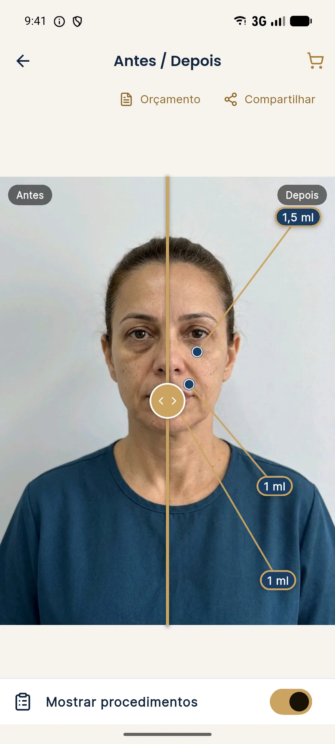
How to use the magnifier in the conversation is covered in presenting the treated area with the magnifier.
Which traps distort the impact ranking?
- Bias toward what is easy to show: areas that "look good" in the photo gain priority for that reason alone.
- Ticket bias: putting the most expensive area first. The ranking needs to be auditable by the patient.
- Trend bias: prioritizing what is in vogue on social media. Unrealistic references are the subject of filter face: how to handle unrealistic references.
- Starting-photo bias: a photo with poor light or a forced expression distorts perceived impact. The standard is in planning photo and lighting.
- Homogenization bias: applying the same ranking to every patient, ignoring proportions and ancestry, as in facial proportions vary between populations.
How does the ranking fit into the plan's timeline?
The ranking becomes a schedule. The first two or three areas form the first stage, and the following ones enter according to the clinical interval and the budget. The logic of distributing over time is in 12-month facial harmonization plan and in what to treat first in facial harmonization. When prioritizing, also remember the financial limit, covered in prioritizing by the patient's budget.
When should you review the ranking?
After the first stage. The face changes, the swelling passes and the patient's perception adjusts. Reordering with the follow-up photos is part of good practice, as in re-planning after the first session. Plans that are too rigid tend to stay faithful to a face that no longer exists.
Frequently asked questions
How do you know which area has the most visual impact?
Combine the patient's concern with a reading of the face: areas that project or support, such as the cheekbone, chin and jawline, usually change the perception of the whole more than refinement areas.
Is the area that bothers the patient most always the one with the greatest impact?
Not always. The bother may lie in a detail whose cause is structural, and treating only the detail produces a result that satisfies little. It is worth investigating the origin first.
Can I start with what the patient wants, even if it is not the greatest impact?
You can, as long as the conversation makes clear what is left out and what that means for the whole. The final decision is the patient's, with information.
Does Harmy help compare impact between areas?
Yes. You can project isolated and combined areas, and the patient sees what each marking changes on their own face, with the ml displayed over the After.
Harmy images are illustrative simulations generated by AI from the patient's photo to support the conversation between practitioner and patient. They are not a promise of results. Indication, technique, product and volume are solely the decision of the licensed practitioner. This content is for information only and does not replace a clinical assessment.