
Asymmetric smile: how to assess it in photo and video

- Muscle
Quick facts
- Concern
- One side of the smile rises more, opens more or shows more teeth
- Method
- Photo and short video of the spontaneous smile and the posed smile
- Origins
- Muscle, bone, teeth, lip or soft tissue
- Caution
- Do not treat before knowing where the asymmetry comes from
The smile is the most-seen expression on the face and one of the hardest to assess, because it is dynamic: it involves muscles, lips, teeth, bones and the person's willingness to show their smile. The patient says "one side goes up more" or "my smile is crooked," and the professional's opening question should be where the asymmetry originates. Without answering that, any procedure becomes trial and error.
This page proposes a simple method: record the smile in photo and video, separate the possible origins and only then decide. For the broader topic of mouth deviation, see mouth off the midline, and for the smile in planning analysis, smile analysis in HOF.
Why is the smile asymmetric?
Almost every smile has some asymmetry. A small difference between the sides is normal and is often part of a person's charm. What deserves attention is a difference that bothers the patient or that changed recently. The origins fall into five groups:
| Origin | How it presents | Clinical note |
|---|---|---|
| Muscular | One side lifts the mouth corner more; difference in strength | Muscles such as the zygomaticus major and the levator anguli oris |
| Dental | Tilted occlusal plane, deviated dental midline | Integrated dental assessment |
| Bony | Asymmetric mandible or maxilla, deviated chin | Photos, palpation and, if needed, imaging |
| Soft tissue | Lip with different volume or contour | May respond to harmonization |
| Neurological or post-trauma | Paresis, scar, surgical sequela | Refer for specific assessment |
The muscular cause is the one most related to HOF, and therefore the one that demands the most care. The modiolus at the corner of the mouth, where several muscles converge, is a key point: any change there alters the entire smile.
How to record the smile
A still photo is not enough. The best record combines:
- Rest: neutral face, from the front, to see the starting point.
- Spontaneous smile: make the patient laugh for real (a light question or a joke) and capture it on short video. A spontaneous smile is usually more symmetric than a posed one.
- Posed smile: what the patient does when they want to show their teeth.
- Maximum smile: with maximum opening and retraction.
- Speaking: pronouncing letters such as "ee," "oo," "oh" to see the dynamics.
Compare the three versions: if the spontaneous smile is symmetric and the posed one is not, there may be a habit or compensation component. If the asymmetry is the same in all of them, it is structural. The discussion of when to photograph with or without a smile is in photo with or without a smile in planning.
What harmonization can do
The approach depends on the origin, and the options are always at the discretion of the licensed professional.
Soft tissue. When one side of the lip has less volume or the mouth corner is more sunken, lip filler and mouth corner filler can balance the contour. This is where harmonization is most predictable, though still with limits.
Muscular. A small amount of toxin can balance the strength of muscles that pull the mouth corner, such as the depressor anguli oris. The indication is delicate, because the same toxin can cause the opposite effect. See toxin in the depressor anguli oris and asymmetric smile after toxin.
Dental. The occlusal plane and midline belong to dentistry. A partnership between HOF and orthodontics, or between HOF and restorative dentistry, is often the only way to treat the real cause. See HOF integrated with dentistry.
Bony. Relevant skeletal asymmetries are a matter for orthodontics and surgery. HOF can camouflage part of the difference in soft tissue, without correcting the bone.
How to talk to the patient
A patient with a smile concern is usually noticing something almost nobody else sees. Validate the observation, show what the record reveals and avoid phrases like "that is normal" without explanation. Also avoid promising symmetry: see how to point out asymmetry without offending.
What Harmy shows and what it does not
Harmy works with a single frontal photo at rest and simulates the marked area. It does not simulate smiling or movement. For this concern, the useful points are Lips and Mouth Corner, which show how a balance of volume can change the outline of the mouth at rest. The projection changes only what was marked, without altering expression.
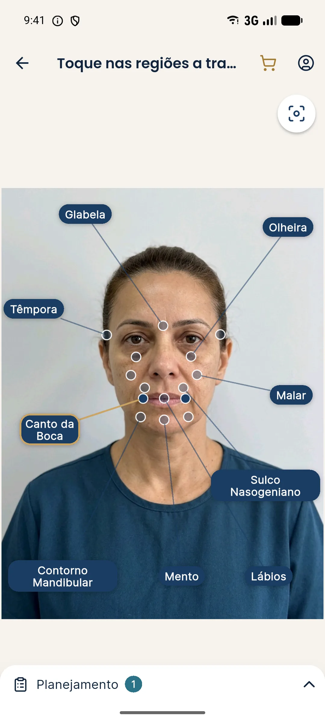
For the design of the lip curve in the smile, see smile arc and lower lip curve and buccal corridor and smile. When a patient arrives with a dull smile but no asymmetry, the page is dull smile. Volumes and technique are left to the discretion of the licensed professional, in line with the rules of their professional council in Brazil.
Frequently asked questions
Why is my smile crooked?
A smile depends on several muscles, the teeth, the bones and the lip. A small difference between the sides is common. The origin can be muscular, dental, bony or soft tissue, and each calls for a different approach.
Can harmonization correct an asymmetric smile?
When the origin is in soft tissue, such as the volume or contour of the lip and mouth corner, partial improvement is possible. When it is muscular or skeletal, the result is usually limited, and assessment defines the path.
Can botulinum toxin improve smile asymmetry?
In some cases, the balance of muscles that pull the mouth down or up can be worked with a small amount of toxin. It can also cause asymmetry if poorly indicated. The decision belongs to the licensed professional.
Does smile asymmetry depend on the teeth?
Often. A tilted occlusal plane, a deviated dental midline and tooth position change how the smile reads, and in that case dental assessment is part of the plan.
Harmy images are illustrative simulations generated by AI from the patient's photo to support the conversation between practitioner and patient. They are not a promise of results. Indication, technique, product and volume are solely the decision of the licensed practitioner. This content is for information only and does not replace a clinical assessment.