
Shadow beside the nose: nasolabial fold and cheekbone
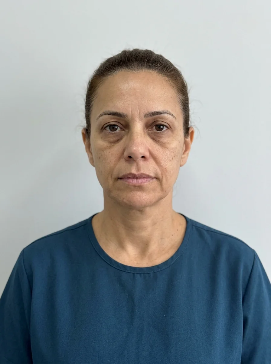
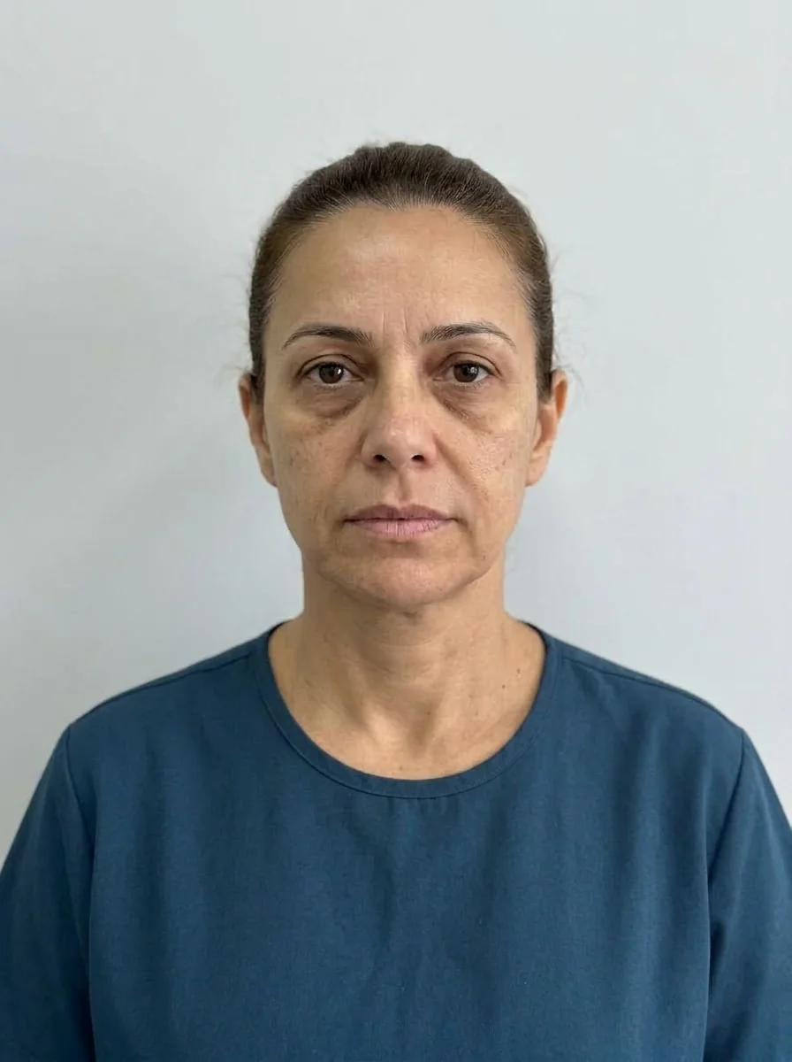 BeforeSimulation
- Highlighted area
Quick facts
- Concern
- Shadow or crease beside the nose that seems to run from the nasal ala down to the mouth
- Structures
- Nasolabial fold, nasolabial compartment, cheekbone and premaxilla
- Pitfall
- Filling the fold on its own can create heavy, artificial volume
- Risk
- High vascular risk area, close to the facial and angular arteries
The person looks in the mirror and sees, on each side of the nose, a shadow running down like a small groove. In photos it gets stronger. Patients call it many things: "smile lines", "nose crease", "fold", "hole beside the nose". The meaning is the same: a marked shadow between the nasal ala and the mouth, which gives a tired, aged look.
The most common approach is to fill the fold, and that is where the greatest risk of error lies. This page shows what makes up the shadow, how to separate what is fold from what is lack of support, and how to order the plan.
What this shadow is made of
The shadow beside the nose has three layers of cause, and they add up.
- The fold itself. The nasolabial fold, known as smile lines, is a natural crease between the side of the nose and the corner of the mouth. It exists in every face and only becomes a concern when it deepens.
- The nasolabial compartment. A fat pad located next to the fold. When it descends, it forms a crease and accentuates the fold. See nasolabial compartment.
- What sits above. Loss of projection in the cheekbone and the paranasal area removes support and lets tissue fall over the fold. See SOOF and premaxilla.
When the patient points to the fold, the professional's eye should move upward. What looks like "lack of volume in the crease" may be "too much descent from above". See treating the fold or the cause of the fold and nasolabial fold and cheekbone: combining them in the plan.
How to assess
Assessment requires looking at the face from the front, in three-quarter view and in motion. The fold changes appearance when smiling.
Comparing with younger photos of the patient shows how much of the fold is constitutional and how much is acquired. People who always had a marked fold respond differently from those who developed it with volume loss. See also nasolabial fold different on one side and deep fold from nose to mouth.
Fill the fold or support the cheekbone
The central discussion of this concern is the order of treatment.
| Approach | When it makes sense | Risk of error |
|---|---|---|
| Support the cheekbone first | The lift test improves the fold | Excess lateral volume |
| Fill the fold first | Deep fold with an adequate cheekbone | Heavy volume and a "full" crease |
| Combine both | Volume loss and an accentuated fold | Too much product |
| Skin quality approaches | Laxity and thin skin | Expectation of a lift |
The delicate point is the second: filling the fold without support above produces a stuffed crease that weighs down the midface and increases descent. In many patients, cheekbone filler softens the fold indirectly and more naturally. When the fold still bothers the patient, nasolabial fold filler comes next, in a conservative amount.
Other options: collagen biostimulator in selected cases and thread lift, each with its own indications, risks and limits. See biostimulator in the nasolabial fold and PDO threads in the nasolabial fold.
The vascular risk of the area
Few areas of the face demand as much anatomical knowledge as this one. The facial artery and the angular artery run through the area near the fold, and the angular artery communicates with the circulation of the eye. Vascular occlusion, although rare, can cause skin necrosis and, in extreme cases, vision loss.
For this reason, the area calls for a qualified professional with specific training, hyaluronidase on hand, and knowledge of the warning signs. Intense pain during injection, skin color change and visual changes are emergency signs. See vascular occlusion in the nasolabial fold, visual complication after filler and vascular occlusion.
Timeline of a plan
The waiting time between stages is decisive. Swelling in the cheekbone can hide the improvement of the fold, and reassessing too early leads to unnecessary additions.
How Harmy fits in
Harmy has the Cheekbone and Nasolabial Fold points, and the projection shows the effect of volume on the frontal photo. It is a useful resource for this concern, because the patient can see the difference between working on the fold and working on the cheekbone.
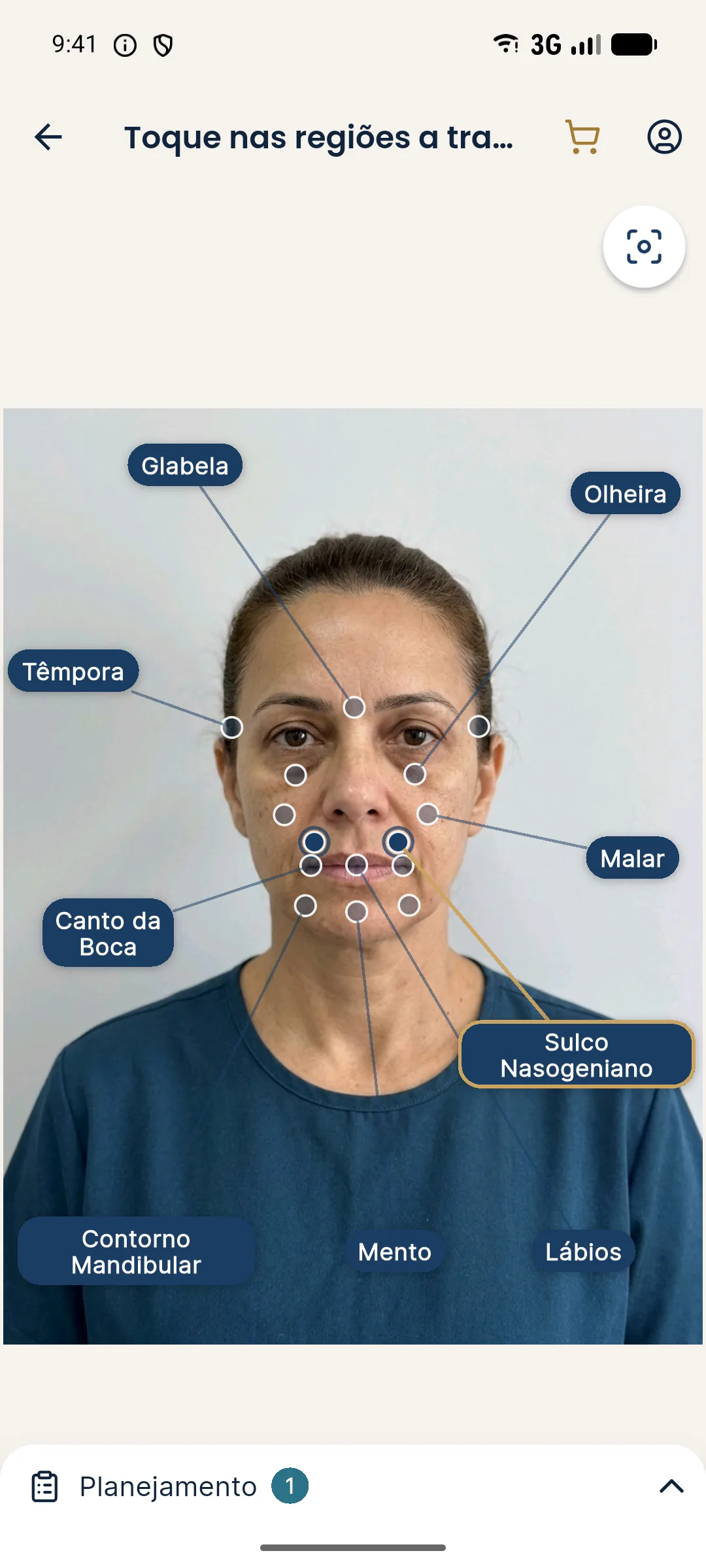
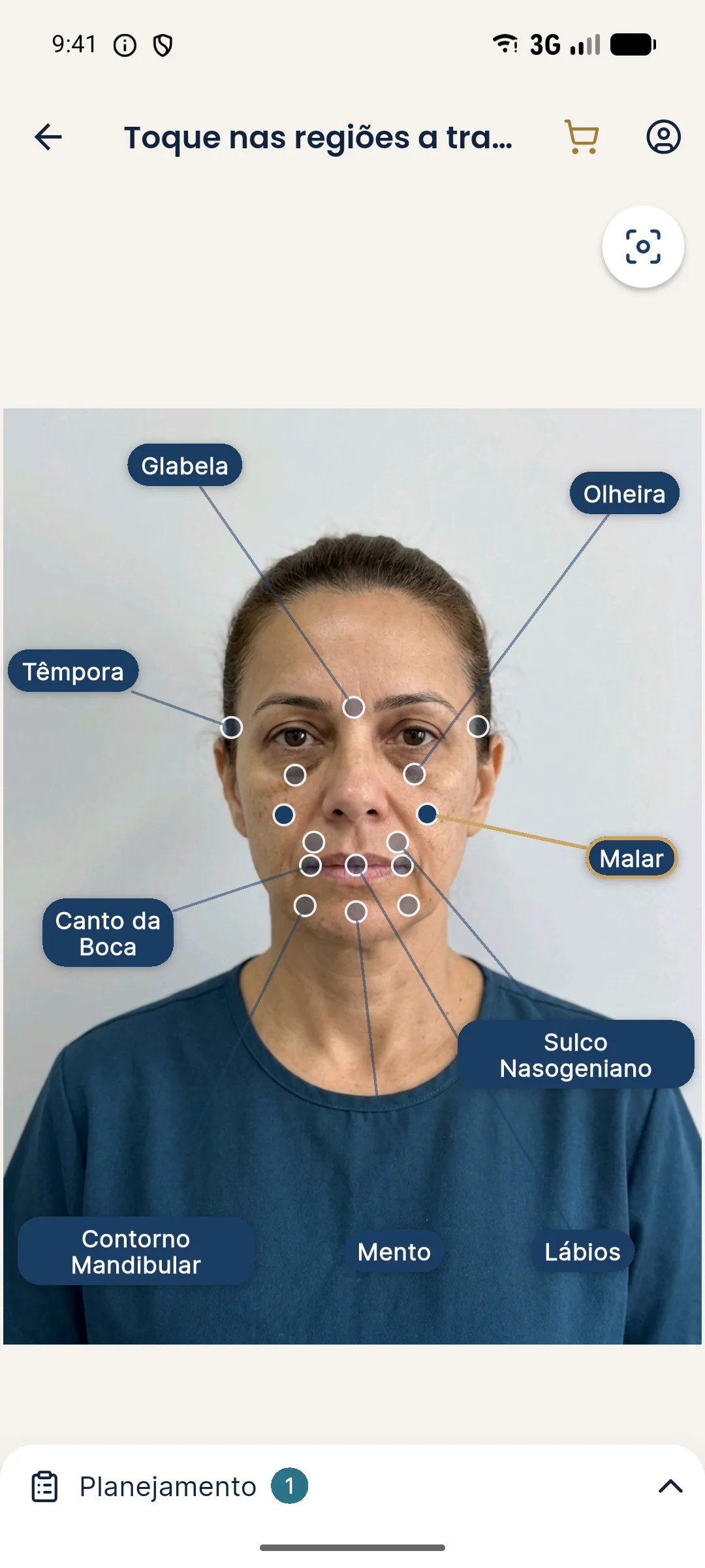
This comparison is the most honest way to justify a plan that starts with the cheekbone: the patient sees the logic on their own face and more readily accepts starting with a point that is not the one they pointed to.
Record keeping
Record the lift test, the photos, the staged plan and the explanations about risk. Volumes and techniques are at the discretion of the qualified professional, after assessment and in line with the rules of their professional council in Brazil.
Frequently asked questions
What is the difference between the shadow beside the nose and the nasolabial fold?
The nasolabial fold is the crease between the side of the nose and the corner of the mouth. The side shadow is the visual reading of that crease plus the depth of the cheekbone and the paranasal area.
Is filling the fold the best treatment?
Not always. In many cases, improving support of the cheekbone and midface softens the fold indirectly and more naturally. The decision belongs to the professional, after assessment.
Does the premaxilla influence this shadow?
Yes. A lack of projection in the area beside the nasal opening can deepen the shadow and close the angle between the nose and the upper lip.
Is there risk in treating this area?
Yes. The facial and angular arteries run close by, and vascular occlusion, although rare, is serious. Hyaluronidase must be available whenever hyaluronic acid is used.
Harmy images are illustrative simulations generated by AI from the patient's photo to support the conversation between practitioner and patient. They are not a promise of results. Indication, technique, product and volume are solely the decision of the licensed practitioner. This content is for information only and does not replace a clinical assessment.