
Asymmetric chin: how to assess chin deviation
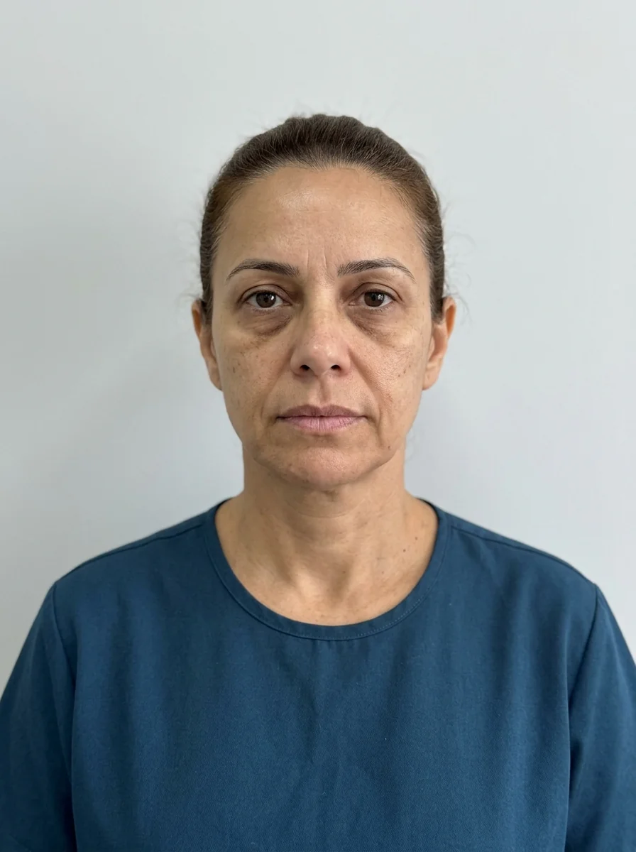
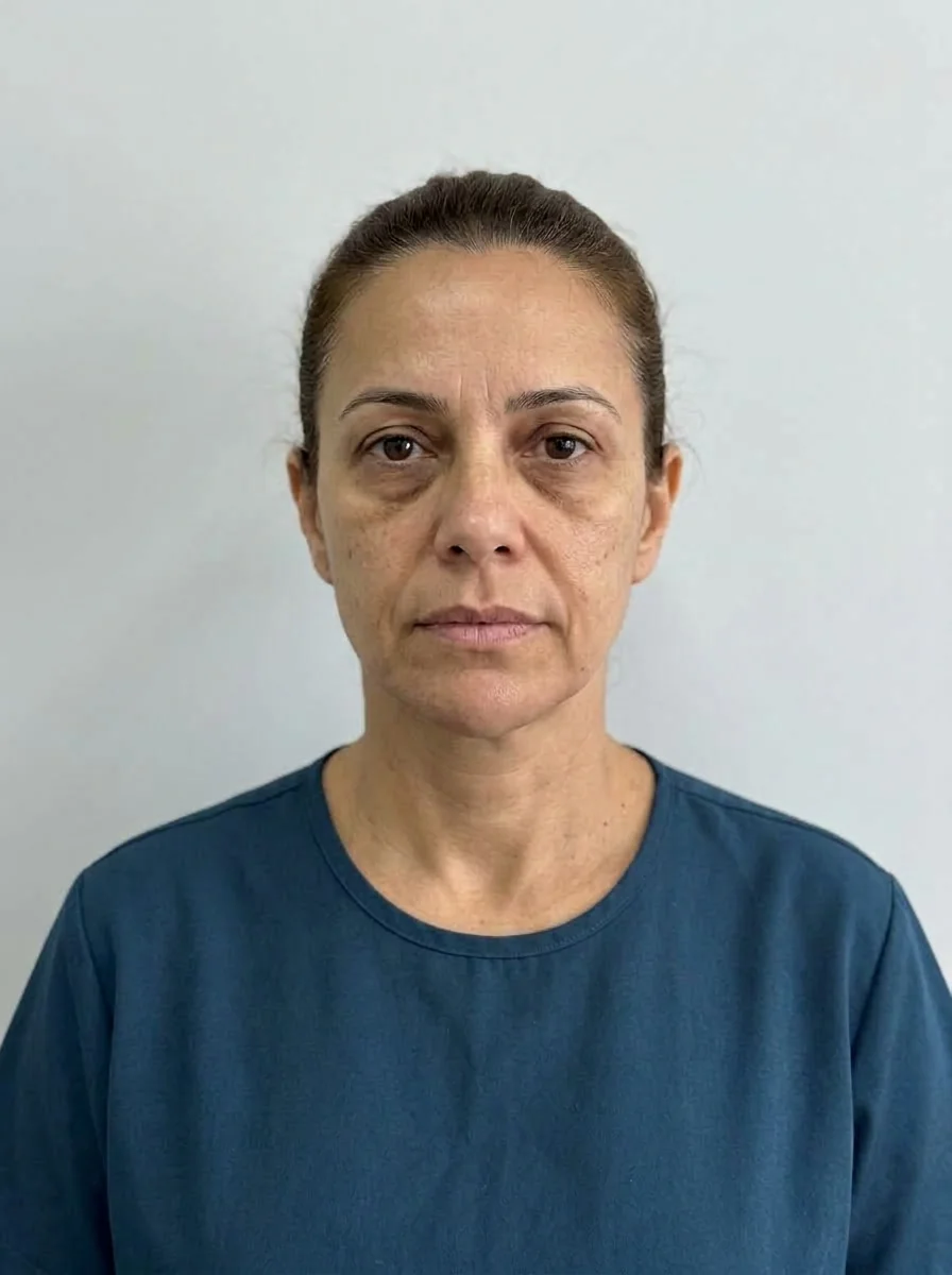 BeforeSimulation
- Nerve
- Highlighted area
Quick facts
- Concern
- A chin that looks crooked, shifted to one side, or fuller on one side
- Key reading
- Distinguish bone deviation, muscle asymmetry, volume, and bite before treating
- Supporting measure
- Front photo with midline, pogonion, and comparison of the height of the two mandibular angles
- Limit
- Volume camouflages mild differences, but does not correct a skeletal or bite deviation
The patient looks at the photo and says the chin is "crooked." Sometimes it is a difference of a few millimeters that only they see. Other times, the deviation is clear and follows the mouth, the jaw, and the bite. The clinical question is the same in both cases: where does the difference come from?
This page organizes the assessment of an asymmetric chin into three origins (bone, muscle, volume) and shows what HOF can do in each. For chin deviation relative to the center of the face, see chin off the midline, and for the bigger picture, asymmetric jaw.
What are the three origins of chin asymmetry?
The same "crooked chin" look can come from very different causes. Separating these causes completely changes the approach.
| Origin | What happens | Clue in the assessment | Role of HOF |
|---|---|---|---|
| Bony | The pogonion is shifted or one side of the jaw is larger | The deviation does not change with expression and follows the bone | Partial camouflage; refer if marked |
| Muscular | One side of the mentalis or depressor muscle works more | The asymmetry shows when speaking, smiling, or puckering the chin | Toxin, at the professional's discretion |
| Volume | One side has less fat or soft tissue | Visible difference at rest, without bone deviation | Conservative filler |
| Occlusal | The bite pushes the jaw to one side | The dental midline does not match; deviation on closing | Orthodontics; HOF as a complement |
In many patients the origins are mixed. The anatomy involved is that of the pogonion, the mentalis muscles, and the mandible, and it is useful to review these structures before deciding.
How to take photos for assessment?
A standardized front photo is the basic tool. Ask the patient to hold the head straight (Frankfort plane horizontal), lips relaxed, and teeth in light occlusion. Adjust the light to avoid shadows that change the contour. Then draw reference lines on the screen:
- The facial midline, passing between the eyebrows, through the tip of the nose and the philtrum.
- The position of the pogonion relative to that line.
- The height of the two mandibular angles, with a horizontal line.
- The tilt of the mouth line and of the eyes.
If the chin is shifted to one side relative to the philtrum, but the philtrum is at the center, the deviation is of the chin. If the philtrum also deviates, the asymmetry is broader. Reading the midline is covered in facial midline and in midline deviation: how to plan.
How do you tell bone, muscle, and volume apart in practice?
A few simple tests help, without replacing a specialist exam. Ask the patient to fully relax the face and compare. Then ask them to smile, speak, and close the mouth tightly. If the asymmetry increases with movement, there is a muscular component. If it stays identical at rest, the factor is structural.
Palpating the area also guides you. The bone contour is usually palpable, and a difference in projection that does not change with gentle pressure suggests a bony origin. A difference that "disappears" when the soft tissue is compressed indicates a volume component. The conclusion is the examiner's, and uncertainty justifies referral.
What can harmonization do?
Within what the technique allows, the options vary by origin:
- Mild volume asymmetry: chin filler with different volume on each side, conservatively, at the discretion of the licensed professional. See chin filler.
- Muscular component: toxin in the mentalis muscle when there is asymmetric overactivity. See mentalis toxin.
- Jawline: when the difference extends to the angle, the plan may involve the mandible. See chin and jaw: combining in the plan.
- Bone or bite asymmetry: partial camouflage and referral, when the difference is larger.
In the chin area, the risks include bruising, swelling, asymmetry, and nodules. There are vascular and nerve structures in the area, such as the mental nerve, and hyaluronidase must be available when hyaluronic acid is used. Reinforce with the patient that the correction may be partial.
How to explain it to the patient
The conversation needs to make clear what is possible. Many patients believe symmetry is solved with one "injection," and that belief creates dissatisfaction. Show the photo with the reference lines, point out where the difference is, and say which part you can act on.
To handle asymmetry without offending, see how to point out asymmetry without offending, and for aligning expectations, aligning expectations about asymmetry.
And Harmy in this case?
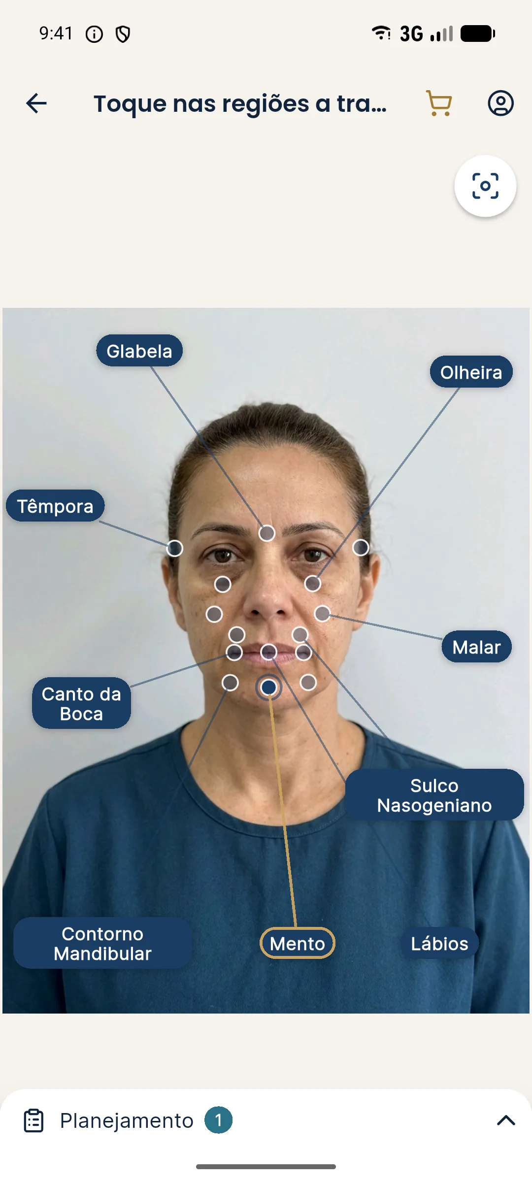
The projection in Harmy uses only the front photo and shows the volume you mark on the Chin point. That helps visualize the effect of volume replacement, but the app treats the Chin point as a single area, with one total ml, and does not set different volumes on each side. For asymmetry, the simulation serves as an illustration of the direction of the adjustment, not as a fine map of the distribution.
When to refer?
Some situations call for another specialty's assessment before or alongside HOF:
- Marked chin deviation accompanied by a bite deviation.
- Asymmetry that recently got worse with no clear cause, which may call for medical investigation.
- Pain or limited mouth opening, which suggest a joint problem.
- History of trauma or jaw surgery with a persistent complaint.
- The patient expects complete symmetry.
Orthodontic and surgical guidance can change the picture completely. See orthodontics and profile change and referring patients in HOF.
Records
Save the photo at rest, smiling, and with the mouth closed, the reference lines, the probable classification (bony, muscular, volume, mixed), the plan in ranges, and the guidance about limits. Compare at follow-up with the same angle and light. The history per patient in Harmy helps keep sessions side by side, and standardized clinical photography describes the method.
Frequently asked questions
Can an asymmetric chin be fixed with filler?
When the difference is in volume or a mild contour, filler can balance the appearance. If the deviation comes from the bone or the bite, the effect is partial camouflage, and the professional should explain the limit.
Does everyone have some degree of chin asymmetry?
Most people have some facial asymmetry. The concern becomes clinical when the difference bothers the patient or is visible relative to the whole face.
How do I know if the chin deviation is bony?
Clinical assessment, tooth position and, when needed, imaging help. If a skeletal deviation is suspected, referral to a specialist is the next step.
Can toxin help with chin asymmetry?
When there is overactivity of the mentalis muscle on one side, toxin can soften the difference in movement. It does not correct bone or volume, and the decision belongs to the licensed professional after assessment.
Harmy images are illustrative simulations generated by AI from the patient's photo to support the conversation between practitioner and patient. They are not a promise of results. Indication, technique, product and volume are solely the decision of the licensed practitioner. This content is for information only and does not replace a clinical assessment.