
Disproportionate Midface: Assessment
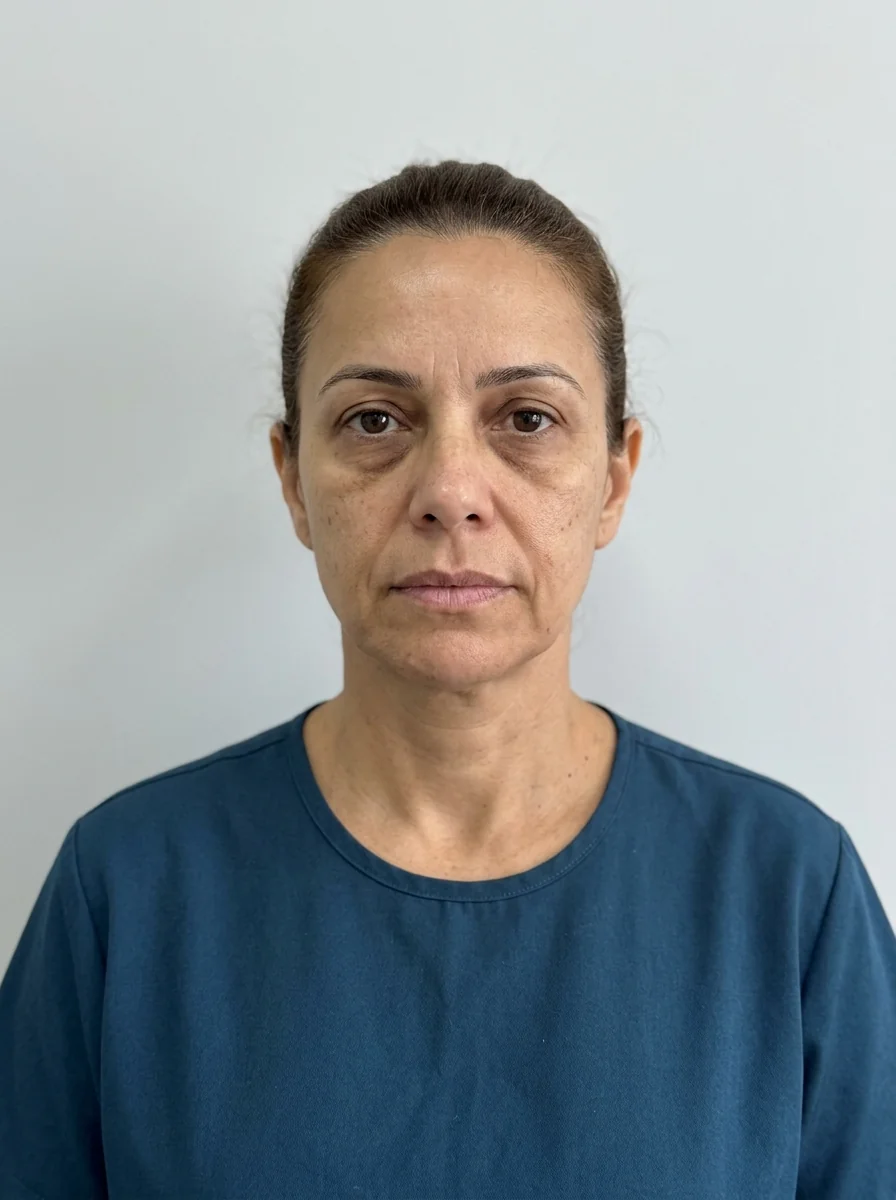
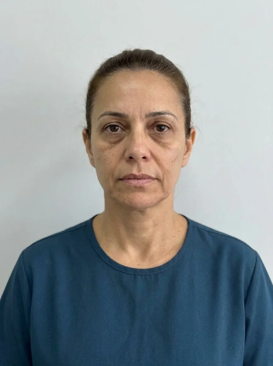 BeforeSimulation
- Nerve
- Highlighted area
Quick facts
- Concern
- Middle of the face that looks too big, flat, elongated or out of proportion with the forehead and chin
- Key question
- What dominates: the nose, the cheekbone, the maxilla or the cheek fat?
- Assessment stance
- Look from the front, in profile and in three-quarter view, because the midface looks different in every view
- Plan risk
- Enlarging the cheekbone in a face that already looks wide in the middle, or ignoring the nose that dominates the third
"The middle of my face is too big", "my face is flat in the middle", "my nose dominates everything". The phrases are different, but they describe the same territory, the midface, where the nose, cheekbone, maxilla and cheek live. It is a third that changes appearance a lot depending on the view (front, profile, three-quarter), and for that reason it is easily misjudged from a single photo.
This page organizes the investigation of midface disproportion, which can be by excess, by deficit or by mismatch with the other thirds. For the case of a profile without projection, see profile without midface projection, and for tissue descent, midface ptosis.
What makes up the midface
The midface runs from the glabella (or the brow line) to the subnasale. It contains the nose, the eyes and periorbital area, the cheekbone and the zygomatic bone, the maxilla and the cheeks. It is the third that most defines how young and full the face looks, and also the one most affected by tissue descent over the years.
Because of this complexity, "disproportionate midface" says little without a follow-up question: disproportionate in what dimension (height, width, projection) and because of which structure?
Three types of disproportion
| Type | How it presents | Note |
|---|---|---|
| By excess | Long or wide nose, full cheek, projected maxilla | HOF has a limited role; the conversation about adjusting perception is central |
| By deficit | Malar hypoplasia, recessed maxilla, volume loss | Volume replacement is the usual conversation |
| By mismatch | Normal midface, but upper and lower thirds out of scale | Treating the midface may not be the answer |
The third by excess is the least favorable for HOF. Reducing is not what injectables do best, and the professional needs to say so. The third by deficit is the closest to what HOF does, but it requires assessing the skeleton before adding volume. The third by mismatch is a reminder that the plan starts with the whole.
The nose as the dominant factor
In many patients, the complaint is about the face, but the reason is the nose. A projected or long nose dominates the third, and the rest looks "small" around it. The nasal index, the alar width and the nasal projection help make this objective. HOF has a limited role in reducing the nose, but nonsurgical rhinoplasty appears as an option to adjust lines and the tip in selected cases, with specific risks and in line with the rules of the professional's board. Read explaining nonsurgical rhinoplasty to the patient.
When the nose dominates, reinforcing the cheekbone can balance the reading, and occasionally it is the only intervention needed. But the decision requires seeing the profile and the three-quarter view, not just the front.
The maxilla and the bone
The maxilla and the zygomatic bone support the midface. When the skeleton is poorly projected, volume placed over it compensates in part, but does not replace the support. Faces with a recessed maxilla tend to have a flat midface, and a profile assessment shows this clearly. The topic connects with maxillary projection and midface in profile.
If there are signs of maxillary deficiency or skeletal class III, a conversation with an orthodontist or maxillofacial surgeon is part of the care, and HOF acts as a complement.
Fat and aging
Over time, midface fat descends, the cheekbone loses support and the cheeks change position. A midface that looked proportionate at 30 can look hollow above and heavy below at 50. The complaint becomes "disproportion", but the cause is aging. For the layered reasoning, see aging of the midface and midface fat compartments in the plan.
When there is a real excess of cheek fat, the conversation turns to procedures outside the scope of HOF. The discussion of midface disproportion by excess details this.
Options that enter the conversation
Hyaluronic acid is used to replace volume in the cheekbone and zygomatic area, at the professional's discretion, after assessment. The collagen biostimulator appears in profiles with loss of support and quality, with a gradual response. Threads are discussed in selected cases. To read the limits of each, see biostimulator in the midface, PDO threads in the midface and the procedure cheekbone filler.
How to organize the plan
- Photograph from the front, in profile and in three-quarter view with standardization.
- Measure the height and width of the midface relative to the others.
- Classify the type of disproportion: excess, deficit or mismatch.
- Assess the nose, cheekbone, maxilla and fat as contributors.
- Choose the smallest adjustment that addresses the concern, in steps.
- Reassess with comparison photos and talk about what changed.
Safety
The midface concentrates risk areas: the cheekbone and zygomatic area have the transverse facial artery and branches of the infraorbital nerve; the tear trough and nasolabial fold have branches of the facial and angular arteries; the nose carries a high vascular risk. Complications include swelling, bruising, asymmetry, nodules and, rarely, vascular occlusion. Keep hyaluronidase available and know the warning signs.
How to use Harmy in this conversation
Harmy's projection adds volume at the marked points from a frontal photo. For the midface, the Cheekbone point lets you show the effect of replacement. The app does not show the profile, does not simulate changes to the nose and does not represent reduction, and the patient needs to be told this before the slider is opened.
Questions to test your reasoning
What to record in the patient record
Record the three views, the measurements, the type of disproportion and the dominant cause you identified. Note the conversation about limits, any referrals made and the specific consent. For photos, see standardized clinical photography. For the complete planning approach, complete midface in the plan.
Common mistakes
The first mistake is assessing the midface only from the front. The second is treating the cheekbone without looking at the nose and maxilla. The third is promising the patient that volume will solve what belongs to the skeleton or to excess soft tissue.
Summary for the decision
A disproportionate midface is a concern about the whole, with three types of origin: excess, deficit and mismatch. Photograph in three views, measure, classify, assess the nose, cheekbone, maxilla and fat, and choose the smallest intervention that addresses the concern. HOF reinforces, it does not reduce. Keep hyaluronidase available, document, and act at the discretion of the qualified professional, in line with the rules of their professional board, without promising results.
Frequently asked questions
How can you tell whether the midface is disproportionate?
It is measured on a standardized photo and compared with the upper and lower thirds and with the width of the face. Perception also depends on the nose, cheekbone, maxilla and the volume of the cheek.
Can the nose be the cause of a midface that looks large?
Yes. A long, wide or highly projected nose dominates the third and makes the whole look bigger. The assessment includes the nose and a conversation about what HOF can or cannot adjust.
Does a flat midface improve with cheekbone filler?
It can improve apparent projection when the deficit is one of volume. If the problem lies in the maxilla or bone, the gain is partial and should be explained to the patient.
Can a large midface be reduced?
Reducing bone structure is not the role of HOF. What is discussed is adjusting perception, always at the professional's discretion after assessment, with realistic expectations.
Harmy images are illustrative simulations generated by AI from the patient's photo to support the conversation between practitioner and patient. They are not a promise of results. Indication, technique, product and volume are solely the decision of the licensed practitioner. This content is for information only and does not replace a clinical assessment.