
Zygomatic and lateral malar: the ogee curve in your HOF plan
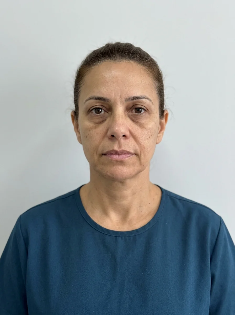
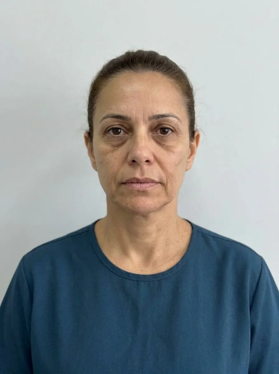 BeforeSimulation
- Nerve
Quick facts
- Concept
- The ogee curve is the S-shaped line running from the temple to the cheek in three-quarter view
- Areas involved
- Zygomatic, lateral and anterior malar, zygomatic arch and temple
- Reading
- Only seen well in three-quarter view, with soft side lighting
- Simulation in Harmy
- Cheekbone point on the frontal photo; the app does not show the three-quarter curve
- Who decides
- Qualified professional, after assessment
Few readings of the face depend as much on angle as the ogee curve. From the front, the malar area looks like a single elevation; in three-quarter view, it forms an S-shaped line that rises along the temple, drops along the zygomatic arch and rounds off at the most projected point of the cheek. This is the line that tends to be lost with aging, and what patients call "sagging cheeks" or a "tired face". Planning the zygomatic and lateral malar areas is, first of all, planning the continuity of this curve. This page helps you read the line, decide where it breaks and talk it through with the patient, without a technique protocol.
What is the ogee curve?
Ogee is a word of architectural origin for a line with two opposite curves, one convex and one concave, forming an S. On the face, it describes the smooth transition between the temple (slightly concave), the zygomatic arch (the bridge) and the malar area (convex). When the curve is continuous, the midface tends to read as youthful and harmonious. When there is a step, flattening or hollowing, it reads as a tired face or "sagging cheeks". See the concept in ogee curve and the bone anatomy in zygomatic arch.
The curve also varies between people, by age, sex, ancestry and bone structure. Not every face has a pronounced ogee, and not every face needs one. The reading is a tool for the exam, not a universal aesthetic target. For the diversity of proportions, see facial proportions vary between populations.
Where does the curve usually break?
There are three common break points, each with its own logic:
| Break point | What you see | Planning question |
|---|---|---|
| Hollow temple | A step at the top of the curve, tired look | Does the temple need support? See temples and malar |
| Poorly defined zygomatic arch | The bridge disappears, the face looks "round" | What is bone and what is soft tissue? |
| Flattened anterior malar | The cheek loses projection, the tear trough and fold get worse | Does the midface support need volume? |
Each point has a different cause and a different possible approach. The reading of ideal projection is in ideal malar projection, and the relationship with fat compartments is in midface fat compartments in the plan. For the history of lines used in assessment, see Hinderer lines in malar assessment.
Why does a smooth transition between points matter?
Because the eye follows the line, not the point. If the plan treats the anterior malar and forgets the lateral malar, or if the temple receives volume unrelated to the zygomatic area, the result is a series of bumps instead of a curve. The patient cannot name it, but notices: "it looks different, but not nice".
The practical approach is to plan the whole as one line and size each point in relation to its neighbor. More projected points need a longer transition, and subtler points can stand alone. But the general rule is to test continuity on the three-quarter photo before and after each stage. See domino effect between facial areas.
Risks that call for attention
The zygomatic and lateral malar areas are close to important vascular and nerve branches. The temple area, in particular, demands detailed anatomical knowledge. Risks include swelling, bruising, asymmetry, nodules, irregularities and, rarely but seriously, vascular occlusion. There is also the aesthetic risk of an "overfilled" look when volumes are added without looking at the curve.
When hyaluronic acid is used, hyaluronidase must be available on site, and the team must recognize warning signs. The anatomy review is in temporal anatomy for injectables and vascular danger zones of the face.
When is bone the issue?
In many faces, the absence of an ogee comes from structure: the zygomatic arch is barely projected, the malar bone is low or the maxilla is retruded. HOF with hyaluronic acid can compensate for part of this, but it does not replace surgery or orthognathic treatment. Saying this clearly is part of informed consent. See when bone is the problem, not volume and facial skeleton before injecting.
It is also worth remembering that facial bone resorption over the years changes the base of the curve. Plans for older patients should take this into account. See facial bone resorption and planning.
How does Harmy fit into this conversation?
In Harmy, the Cheekbone point is marked on the frontal photo, with ml (from 0.5 to 5, in steps of 0.5) and intensity (Subtle, Moderate or Pronounced). For a bilateral area, the ml is the total, split between both sides. The AI respects the volume and changes only what was marked.
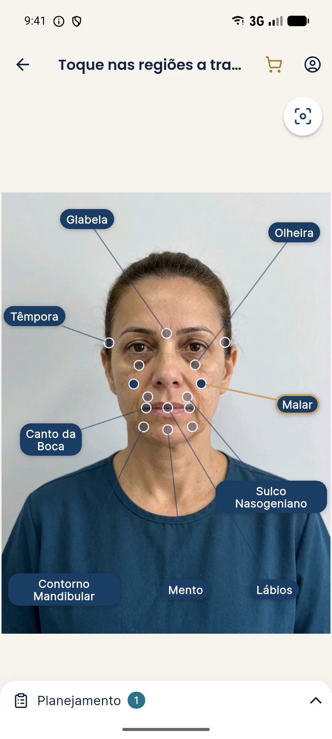
The limitation here is central: the ogee curve can only be read well in three-quarter view, and Harmy uses only the frontal photo. It does not show the S-shaped transition, the zygomatic arch or the bone structure. The honest use is to show the patient that a conservative ml adjustment of the malar area changes how the midface reads from the front, and to explain that the continuity of the curve is assessed during the exam, from another angle.
To understand what the image shows and what it does not, read limits of 2D simulation; to talk it through with the patient, explaining that a simulation is not a guarantee.
How do you organize the plan in stages?
The cadence is in reassessment at 3, 6 and 12 months and the logic of splitting in when to split volume across sessions.
What should you tell the patient?
If the patient brings a reference photo with a very pronounced curve, go back to what their face allows and to facial identity. See facial identity: how to preserve it.
Check before the plan
Quick review
Summary
The ogee curve is the S-shaped line linking the temple to the malar area, and the zygomatic and lateral malar plan is a plan for continuity, not for isolated volume. Read the face in three-quarter view, identify where the line breaks, treat with conservative ml in stages, respect vascular risk and use Harmy as support for the conversation, knowing it uses only the frontal photo.
Frequently asked questions
What is the ogee curve in the face?
It is an S-shaped line that, seen in three-quarter view, runs from the temple to the malar area, passing through the zygomatic arch. It is usually associated with a youthful, smooth midface.
Do I need to treat the zygomatic and lateral malar areas together?
Not always. Many faces need just one point. What matters is the continuity of the curve: isolated, abrupt points tend to break the transition.
Can I assess the ogee from the frontal photo alone?
It is not ideal. The curve is best seen in three-quarter view, with soft side lighting. The frontal photo shows projection, but not the S-shaped transition.
Does Harmy simulate the ogee curve?
No. Harmy uses only the frontal photo and simulates the ml of the Cheekbone point. The three-quarter curve has to be assessed during the exam.
Harmy images are illustrative simulations generated by AI from the patient's photo to support the conversation between practitioner and patient. They are not a promise of results. Indication, technique, product and volume are solely the decision of the licensed practitioner. This content is for information only and does not replace a clinical assessment.